. 2010 Jul 16;400(3):393-412.
doi: 10.1016/j.jmb.2010.04.059. Epub 2010 May 4.

Regulation of PKR by HCV IRES RNA: importance of domain II and NS5A

Affiliations

Regulation of PKR by HCV IRES RNA: importance of domain II and NS5A

Rebecca Toroney et al. J Mol Biol. .

Abstract

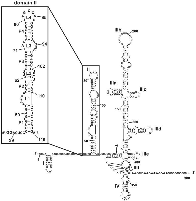
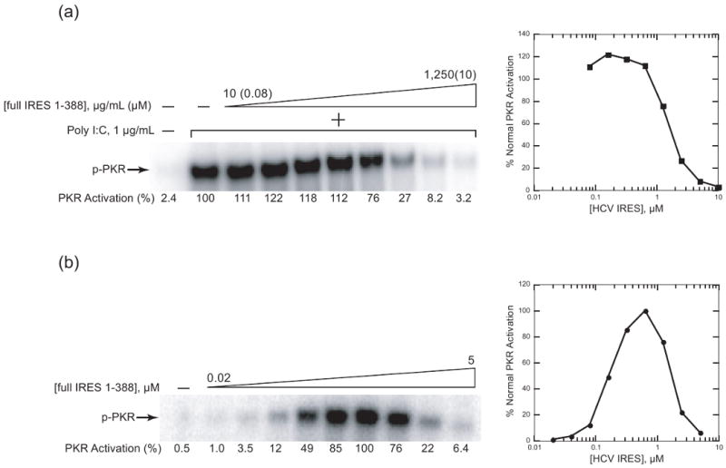
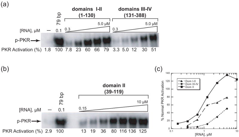
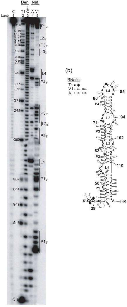
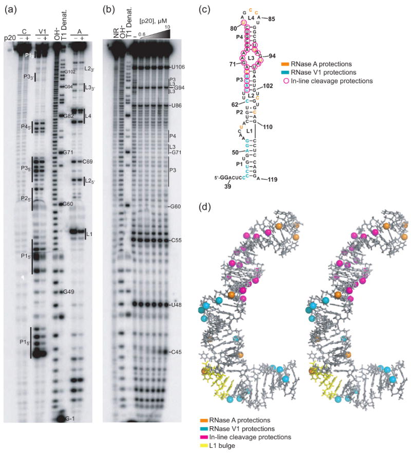
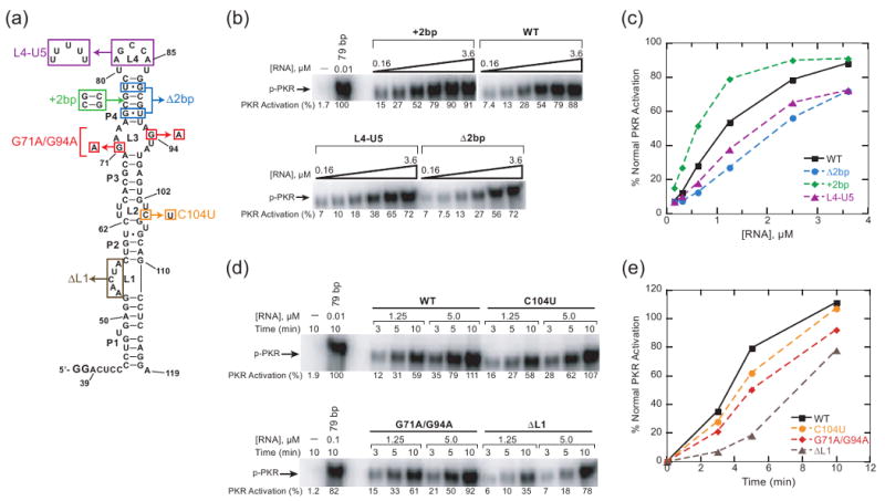
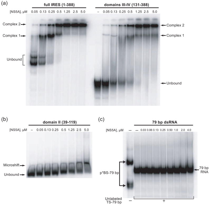
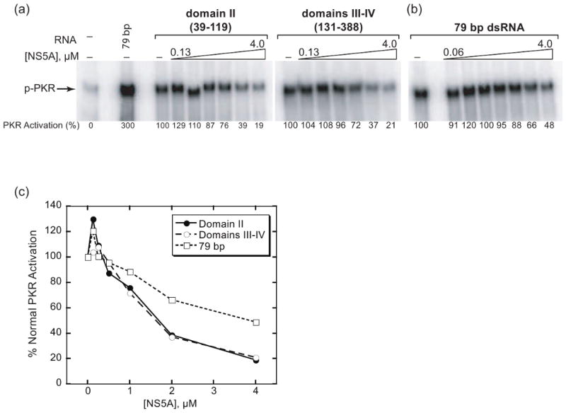
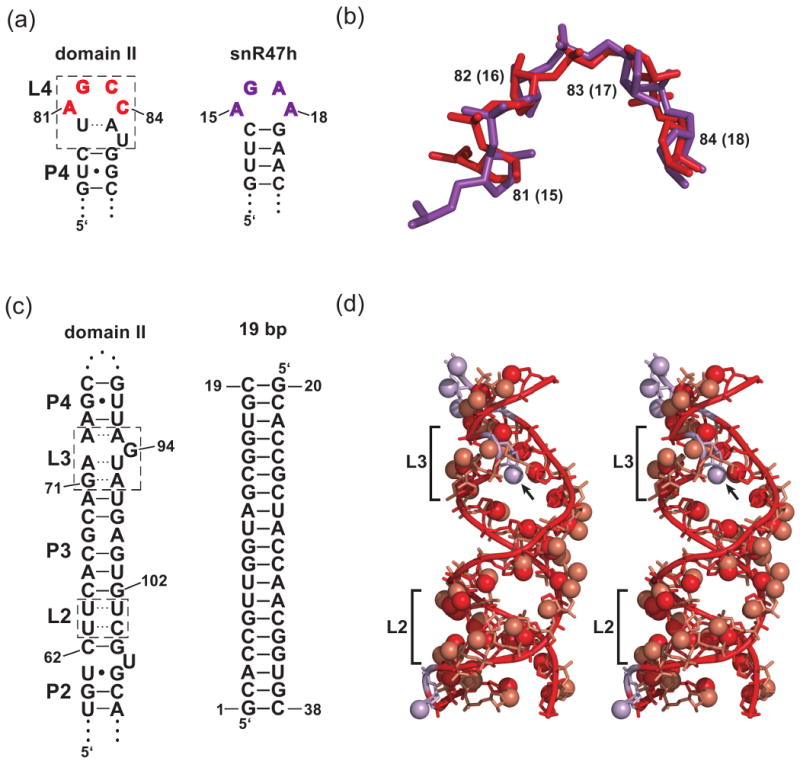
Protein kinase R (PKR) is an essential component of the innate immune response. In the presence of double-stranded RNA (dsRNA), PKR is autophosphorylated, which enables it to phosphorylate its substrate, eukaryotic initiation factor 2alpha, leading to translation cessation. Typical activators of PKR are long dsRNAs produced during viral infection, although certain other RNAs can also activate. A recent study indicated that full-length internal ribosome entry site (IRES), present in the 5'-untranslated region of hepatitis C virus (HCV) RNA, inhibits PKR, while another showed that it activates. We show here that both activation and inhibition by full-length IRES are possible. The HCV IRES has a complex secondary structure comprising four domains. While it has been demonstrated that domains III-IV activate PKR, we report here that domain II of the IRES also potently activates. Structure mapping and mutational analysis of domain II indicate that while the double-stranded regions of the RNA are important for activation, loop regions contribute as well. Structural comparison reveals that domain II has multiple, non-Watson-Crick features that mimic A-form dsRNA. The canonical and noncanonical features of domain II cumulate to a total of approximately 33 unbranched base pairs, the minimum length of dsRNA required for PKR activation. These results provide further insight into the structural basis of PKR activation by a diverse array of RNA structural motifs that deviate from the long helical stretches found in traditional PKR activators. Activation of PKR by domain II of the HCV IRES has implications for the innate immune response when the other domains of the IRES may be inaccessible. We also study the ability of the HCV nonstructural protein 5A (NS5A) to bind various domains of the IRES and alter activation. A model is presented for how domain II of the IRES and NS5A operate to control host and viral translation during HCV infection.


Figures

Fig. 1
Fig. 1
Secondary structure of 5′-end of HCV RNA., Shown are nucleotides 1-388, which we refer to as ‘full-length HCV IRES RNA’. Domains are numbered in Roman numerals, with domain II boxed. Translation start codon is boxed (domain IV). Inset: Secondary structure of domain II construct employed and determined in this study containing residues 39-119, with the two G nucleotides added at the 5′-end to aid in transcription priming in bold. Pairing (P1, P2…) and loop regions (L1, L2…) in domain II are labeled. Asterisk downstream of domain II indicates the 3′-end of domains I-II (1-130) construct and the 5′-end of domains III-IV (131-388) construct. Domain III is further labeled according to its subdomains IIIa-IIIf.
Fig. 2
Fig. 2
Inhibition and activation of PKR by full-length HCV IRES RNA (1-388). (a) Inhibition of poly I:C-mediated activation of PKR by HCV IRES. The amount of poly I:C in all lanes was 1 μg/mL, and concentrations of HCV IRES were 0.08, 0.16, 0.32, 0.64, 1.25, 2.5, 5 and 10 μM. No-RNA/no-poly I:C and no-HCV IRES lanes are included. Phosphorylation activities are provided under the gel and were normalized relative to the lane in which only poly I:C was present. (b) Activation of PKR by HCV IRES. RNA concentrations were 0.02, 0.04, 0.08, 0.16, 0.32, 0.64, 1.25, 2.5, and 5 μM. A no RNA lane is included. Phosphorylation activities were normalized to the most reactive lane, 0.64 μM IRES. For both (a) and (b), 10% SDS-PAGE gels are shown, with position of phosphorylated PKR (p-PKR) indicated, and graphical representations of phosphorylation activities as a function of full-length IRES concentration are provided.
Fig. 3
Fig. 3
Activation of PKR by multiple domains of HCV IRES. (a) PKR activation assays of domains I-II (1-130) (left panel) and III-IV (131-388) (right panel). RNA concentrations for both panels were 0.3, 0.6, 1.3, 2.5, and 5.0 μM. (b) PKR activation assay of domain II (39-119) alone. RNA concentrations were 0.15, 0.3, 0.6, 1.3, 2.5, 5 and 10 μM. For both (a) and (b), 10% SDS-PAGE gels are shown, with position of phosphorylated PKR (p-PKR) indicated. A no-RNA lane and 79 bp dsRNA lane are included. Phosphorylation activities are normalized to the 79 bp lane and are noted under the gel. (c) Graphical representation of phosphorylation activities from panels (a) and (b) as a function of RNA concentration.
Fig. 4
Fig. 4
Activation of PKR and phosphorylation of eIF2α by multiple domains of HCV IRES. (a) PKR/eIF2α phosphorylation by full-length (1-388) HCV IRES. For both − and + eIF2α lanes, IRES concentrations were 0.625, 1.25, and 2.5 μM. (b) PKR/eIF2α phosphorylation by domains III-IV (131-388). For both − and + eIF2α lanes, domains III-IV concentrations were 0.625, 1.25, 2.5, 5, and 10 μM. (c) PKR/eIF2α phosphorylation by domain II (39-119). For both − and + eIF2α lanes, domain II concentrations were 0.625, 1.25, 2.5, 5, and 10 μM. For all panels, 10% SDS-PAGE gels are presented, and positions of phosphorylated PKR (p-PKR) and phosphorylated eIF2α (p-eIF2α) are indicated. When present, eIF2α was in 10-fold excess over PKR. PKR activation for all gels was normalized to the 79 bp (-eIF2α) PKR band in panel (c), and eIF2α phosphorylation for all gels was normalized to the 79 bp (+eIF2α) eIF2α band in panel (c). These values are provided under the gel.
Fig. 5
Fig. 5
Ribonuclease structure mapping of domain II. (a) RNA was 5′-end-labeled and subjected to limited nuclease digestion or hydrolysis. Denaturing 12% polyacrylamide gel is shown. Lanes are as follows: C is a control sample (no nuclease), OH is a limited alkaline digest, and T1, A, and V1 are limited digests with ribonucleases specific for single-stranded G, single-stranded C and U, and double stranded regions, respectively. Lanes 2 and 3 were performed under RNA-denaturing conditions (denoted “Den.”), while lanes 4 and 5 were performed under RNA-native conditions (denoted “Nat.”). T1 digestion was also performed under native conditions (not shown). Nucleotides are denoted to the left of the T1 denaturing lane, and pairing and loop regions of domain II are indicated to the right of the gel. (b) Secondary structure of domain II with positions of cleavage by ribonucleases in panel (a) indicated by symbol. Legend is provided and symbol size is proportional to cleavage intensity.
Fig. 6
Fig. 6
Footprinting of p20 onto domain II. (a) Ribonuclease footprinting. RNA was 5′-end-labeled and saturating p20 (10 μM) was added in indicated lanes. This was incubated at room temperature for 30 min followed by digestion with indicated ribonucleases under native conditions. Control lanes were not subjected to nuclease digestion. Denaturing 12% polyacrylamide gel is shown. Lanes are labeled as per Figure 5. Nucleotides are denoted in the T1 denaturing lanes, and regions of domain II are indicated next to sites of cleavage in both the RNase V1 and A digestion lanes. (b) In-line footprinting. Details are as in panel (a) with the following exceptions. RNA was subjected to partial digestion for 40 h at pH 8.3 in the presence of increasing concentrations of p20 (0, 0.625, 1.25, 2.5, 5, 10 μM). NR denotes no reaction. (c) Secondary structure of domain II, with positions of p20-dependent protection from RNase A, RNase V1, and in-line cleavage indicated. Legend is provided in the figure. (d) Stereoview of averaged domain II NMR structure (PDB ID: 1P5P), with positions of p20-dependent protection of 2′-hydroxyls from RNase A, RNase V1, and in-line cleavage indicated. 2′-hydroxyls are depicted as spheres and colored as per the legend provided in the figure. In cases where protection from nuclease and in-line cleavage occurred on the same 2′-hydroxyl (see panel (c)), bases were colored according to nuclease protections for simplicity.
Fig. 7
Fig. 7
Activation of PKR by domain II mutants. (a) Domain II mutants (L4-U5, Δ2bp, +2bp, G71A/G94A, C104U, and ΔL1) superimposed on secondary structure of domain II wild-type (WT). (b) PKR activation assays of P4 and L4 mutants (Δ2bp, +2bp, L4-U5). RNA concentrations for WT and mutants were 0.16, 0.3, 0.6, 1.5, 2.5, and 3.6 μM. Phosphorylation activities for both gels are normalized to 0.01 μM 79 bp in upper gel and are noted under the gel. (c) Graphical representation of phosphorylation activities from panel (b) as a function of RNA concentration. (d) Time course of PKR activation assays for L1, L2, and L3 mutants (ΔL1, C104U, G71A/G94A). Mutants and WT RNA were tested at 1.25 and 5 μM as indicated, and assays were conducted for 3, 5, and 10 min at each RNA concentration. Phosphorylation activities for both gels are normalized to 0.01 μM 79 bp in upper gel and are noted under the gel. (e) Graphical representation of phosphorylation activities from panel (d) as a function of time, at RNA concentration of 5 μM. For both (b) and (d), 10% SDS-PAGE gels are shown, with position of phosphorylated PKR (p-PKR) indicated.
Fig. 8
Fig. 8
Native mobility gel-shift assays of NS5A binding to HCV domains. (a) NS5A binding to full-length IRES and domains III-IV. Trace amounts of radiolabeled RNA were incubated with the indicated concentrations of NS5A and analyzed by 6% native PAGE. (b) NS5A binding to domain II. Assay was conducted as in panel (a). (c) NS5A binding to 79 bp dsRNA. Bottom strand 79 bp was radiolabeled (p*BS-79 bp) and pre-annealed to excess unlabeled top strand RNA in all lanes except the first lane. Assay conducted as in panel (a) and analyzed by 8% native PAGE. Anomalous migration of single-stranded p*BS-79 as two bands is not unexpected, as BS-79 bp has the potential for strong self-structure, supported by migration as a single band on a denaturing gel and indirect evidence from in vivo experiments.
Fig. 9
Fig. 9
Inhibition of PKR activation by NS5A in the presence of domain II, domains III-IV, or 79 bp. (a) Inhibition of domain II- and domains III-IV-mediated activation of PKR by NS5A. Domain II and domains III-IV concentrations were 2.5 μM. NS5A concentrations were 0, 0.125, 0.25, 0.5, 1, 2 and 4 μM. (b) Inhibition of 79 bp dsRNA-mediated activation of PKR by NS5A. The concentration of 79 bp in all lanes was 0.1 μM, and concentrations of NS5A were 0.06, 0.125, 0.5, 1, 2, and 4 μM. For both panels (a) and (b), 10% SDS-PAGE gels are shown, with position of phosphorylated PKR (p-PKR) indicated. Phosphorylation activities were normalized to the lane in which NS5A was absent and are provided under the gels. (c) Graphical representation of the phosphorylation activities in panel (a) as a function of NS5A concentration.
Fig. 10
Fig. 10
Structural comparisons of domain II with AGNN loop and 19 bp dsRNA. (a) Secondary structures of domain II L4 and snR47h RNA apical loop employed for rmsd analysis. Numbering for snR47h is from the reference . At left, domain II structure contains a portion of P4 and L4, depicted in its tetraloop-like form, as described in the text, in which U80 hydrogen bonds with A85 (dotted line), and U86 is extruded from the loop. The boxed region contains all the nucleotides assigned to L4 according to the phylogentically determined secondary structure (see Fig. 1). Loop nucleotides included in the overlay indicated in red. At right, snR47h RNA AGNN tetraloop, with nucleotides included in the overlay indicated in purple. (b) Overlay of sugar-phosphate backbones from panel (a). Domain II L4 nucleotides AGCC are in red and snR47h AGAA tetraloop are in purple. Numbering for domain II (PDB ID: 1P5P) is shown adjacent to ribose sugars, with snR47h (PDB ID: 1T4L) numbering in parenthesis. All atoms shown were included in rmsd calculations. (c) Secondary structures of domain II P2-P4 region and 19 bp dsRNA employed for rmsd analysis. At left are the regions of domain II included in the comparison. The boxed regions contain all the nucleotides assigned to L2 and L3. Pairing within these loops, as described in the text, is denoted by dashed lines. Nucleotides compared in the overlay are described in the Methods section. (d) Stereoview of overlay of sugar-phosphate backbones from panel (c). This structure is sized such that loops and pairing elements approximately align with the secondary structures in panel (c). Lowest energy NMR structure of domain II is used (PDB ID: 1P5O). Domain II truncations are still in red and 19 bp RNA (PDB ID: 1QC0) is in tan. 2′-hydroxyls are represented by spheres. Brackets indicate positions of L2 and L3, and the arrow indicates the 2′-hydroxyl of domain II G94. The nucleotides in domain II that were excluded from the alignment (A74, G75, C76, G94, and U106) are shown in lavender (see Methods). For clarity, nucleotides 20 and 21 of 19 bp RNA are not shown.

Similar articles

Cited by

References

    1. Balachandran S, Roberts PC, Brown LE, Truong H, Pattnaik AK, Archer DR, Barber GN. Essential role for the dsRNA-dependent protein kinase PKR in innate immunity to viral infection. Immunity. 2000;13:129–141. - PubMed
    1. Williams BR. PKR; a sentinel kinase for cellular stress. Oncogene. 1999;18:6112–6120. - PubMed
    1. Samuel CE. Antiviral actions of interferons. Clin Microbiol Rev. 2001;14:778–809. table of contents. - PMC - PubMed
    1. Manche L, Green SR, Schmedt C, Mathews MB. Interactions between double-stranded RNA regulators and the protein kinase DAI. Mol Cell Biol. 1992;12:5238–5248. - PMC - PubMed
    1. Cole JL. Activation of PKR: an open and shut case? Trends Biochem Sci. 2007;32:57–62. - PMC - PubMed

Publication types

LinkOut - more resources