. 2010 Jul;21(7):1103-14.
doi: 10.1681/ASN.2009090984. Epub 2010 May 6.

IgG glycan hydrolysis attenuates ANCA-mediated glomerulonephritis

Affiliations

IgG glycan hydrolysis attenuates ANCA-mediated glomerulonephritis

Mirjan M van Timmeren et al. J Am Soc Nephrol. 2010 Jul.

Abstract

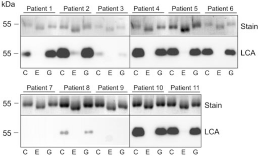
Anti-neutrophil cytoplasmic autoantibodies (ANCA) directed against myeloperoxidase (MPO) and proteinase 3 (Pr3) are considered pathogenic in ANCA-associated necrotizing and crescentic glomerulonephritis (NCGN) and vasculitis. Modulation of ANCA IgG glycosylation may potentially reduce its pathogenicity by abolishing Fc receptor-mediated activation of leukocytes and complement. Here, we investigated whether IgG hydrolysis by the bacterial enzyme endoglycosidase S (EndoS) attenuates ANCA-mediated NCGN. In vitro, treatment of ANCA IgG with EndoS significantly attenuated ANCA-mediated neutrophil activation without affecting antigen-binding capacity. In a mouse model of anti-MPO IgG/LPS-induced NCGN, we induced disease with either unmodified or EndoS-treated (deglycosylated) anti-MPO IgG. In separate experiments, we administered EndoS systemically after disease induction with unmodified anti-MPO IgG. Pretreatment of anti-MPO IgG with EndoS reduced hematuria, leukocyturia, and albuminuria and attenuated both neutrophil influx and formation of glomerular crescents. After inducing disease with unmodified anti-MPO IgG, systemic treatment with EndoS reduced albuminuria and glomerular crescent formation when initiated after 3 but not 24 hours. In conclusion, IgG glycan hydrolysis by EndoS attenuates ANCA-induced neutrophil activation in vitro and prevents induction of anti-MPO IgG/LPS-mediated NCGN in vivo. Systemic treatment with EndoS early after disease induction attenuates the development of disease. Thus, modulation of IgG glycosylation is a promising strategy to interfere with ANCA-mediated inflammatory processes.


Figures

Figure 1.
Figure 1.
EndoS-mediated hydrolysis of the glycan moiety of ANCA IgG. Patient-derived MPO- and Pr3-ANCA IgG are treated with GST-tagged EndoS or GST alone. In the top part (Stain), a Coomassie Brilliant Blue staining is shown of MPO-ANCA IgG (patients 1 through 5) and Pr3-ANCA IgG (patients 6 through 11) that were untreated (C), EndoS-treated (E), or GST control-treated (G). In the bottom part (LCA), an LCA blot is shown from the same ANCA IgGs as presented in the top part. The binding site for LCA is located in the IgG heavy-chain glycan.
Figure 2.
Figure 2.
EndoS-treated ANCA IgG retains antigen-binding capacity. (A, B, D, and E) Using indirect immunofluorescence on ethanol-fixed neutrophils, both deglycosylated (A) and unmodified (B) MPO-ANCA IgG show a perinuclear staining pattern, whereas deglycosylated (D) and unmodified (E) Pr3-ANCA IgG show a cytoplasmic staining pattern. Deglycosylated (○) or unmodified (□) MPO- and Pr3-ANCA IgG samples are serially diluted and tested in a direct ELISA for anti-MPO or anti-Pr3, demonstrating similar titration curves. (C and F) Representative examples of a MPO-ANCA IgG (C) and Pr3-ANCA IgG (F) are shown. Magnification, ×400.
Figure 3.
Figure 3.
EndoS treatment inhibits ANCA-induced neutrophil respiratory burst. (A and B) Examples of a MPO- (A) and Pr3-ANCA IgG-induced (B) respiratory burst in TNF-α–primed neutrophils as measured by the dihydrorhodamine assay. The figure shows neutrophil oxygen radical production in untreated cells (gray) and cells stimulated with deglycosylated (solid line) or unmodified (dotted line) ANCA IgG. (C through F) Summary of the effect of EndoS-mediated deglycosylation of ANCA IgG on MPO-ANCA-induced (n = 5; C and D) and Pr3-ANCA-induced (n = 5; E and F) neutrophil oxygen radical production as determined on neutrophils from two donors and expressed as change in mean fluorescence intensity (donor 1 in C and E, donor 2 in D and F). Bars represent means ± SD. *P < 0.05; **P < 0.01.
Figure 4.
Figure 4.
EndoS treatment inhibits ANCA-induced neutrophil degranulation. (A and B) Effect of EndoS-mediated deglycosylation of MPO-ANCA IgG (■; n = 5) and Pr3-ANCA IgG (□; n = 5) on ANCA IgG–induced lactoferrin release as determined on neutrophils from two donors (donor 1 in A, donor 2 in B). (C and D) Effect of EndoS-mediated deglycosylation of MPO-ANCA IgG (■; n = 5) and Pr3-ANCA IgG (□; n = 5) on ANCA IgG–induced elastase release as determined on neutrophils from two donors (donor 1 in C, donor 2 in D). Bars represent means ± SD. *P < 0.05; **P < 0.01.
Figure 5.
Figure 5.
EndoS-mediated hydrolysis of the glycan moiety of murine anti-MPO IgG does not affect anti-MPO IgG titers after induction of crescentic glomerulonephritis. (A, top) A Coomassie Brilliant Blue staining is shown of murine anti-MPO IgG that was untreated (C), EndoS-treated (E), or GST control-treated (G). (A, bottom) LCA blot is shown from the same samples. (B) Co-administration of anti-MPO IgG and LPS to mice induces NCGN. Mice that received deglycosylated anti-MPO IgG (▴) had similar titers of anti-MPO antibodies compared with mice that received unmodified anti-MPO IgG (□) on day 1. On day 7, anti-MPO titers in both groups (unmodified anti-MPO IgG [▾] and deglycosylated anti-MPO IgG [♦]) are decreased compared with day 1. Data are means ± SD of 12 (day 1) or six (day 7) mice.
Figure 6.
Figure 6.
EndoS-mediated deglycosylation of anti-MPO IgG inhibits induction of hematuria (A), leukocyturia (B), and albuminuria (C) induced by anti-MPO IgG and LPS. (A) Induction of NCGN by unmodified anti-MPO IgG caused marked hematuria after 1 and 7 days (□). Mice that received deglycosylated anti-MPO IgG (▾) had less hematuria after 1 day. (B) Induction of NCGN by unmodified anti-MPO IgG caused increased leukocyturia after 1 and 7 days (□). Mice that received deglycosylated anti-MPO IgG (▾) had mild leukocyturia on day 1, whereas no leukocyturia could be detected on day 7. (C) Albuminuria before (baseline) and at 1 and 7 days after anti-MPO IgG/LPS administration. Mice that received unmodified anti-MPO IgG had increased urinary albumin concentrations on day 1 and even more on day 7. Mice that received deglycosylated anti-MPO IgG had urinary albumin levels that were comparable to baseline levels on days 1 and 7. □, Unmodified anti-MPO IgG; ■, deglycosylated anti-MPO IgG. Bars represent means ± SD. **P < 0.01; ***P < 0.001.
Figure 7.
Figure 7.
EndoS-mediated deglycosylation of anti-MPO IgG reduces early glomerular neutrophil influx. (A and B) Hematoxylin staining (A) and neutrophil staining (B) of a glomerulus on day 1 after disease induction from a mouse that received unmodified anti-MPO IgG and LPS demonstrate marked segmental infiltration of neutrophils. (C and D) Hematoxylin staining (C) and neutrophil staining (D) of a glomerulus on day 1 after disease induction from a mouse that received deglycosylated anti-MPO IgG and LPS demonstrate strongly reduced neutrophil infiltration. (E) Quantification of glomerular neutrophil influx on day 1 after disease induction in mice that received unmodified or deglycosylated anti-MPO IgG. Gcs, glomerular cross-section. ***P < 0.001. Magnification, ×400.
Figure 8.
Figure 8.
EndoS-mediated deglycosylation of anti-MPO IgG prevents development of NCGN induced by anti-MPO IgG and LPS. (A) Overview of renal cortical tissue from a mouse administered an injection of unmodified anti-MPO IgG and LPS 7 days after disease induction, representing the focal and segmental nature of the glomerulonephritis. Glomerular crescents are indicated by arrows. (B) Overview of renal cortical tissue from a mouse administered an injection of deglycosylated anti-MPO IgG and LPS 7 days after disease induction, displaying normal renal morphology. (C) Detail of a glomerulus with a large cellular crescent on day 7, from a mouse that had received unmodified anti-MPO IgG and LPS. (D) Quantification of glomerular crescent formation in mice that received unmodified or deglycosylated anti-MPO IgG expressed as the percentage of glomerular crescents. Horizontal lines represent mean percentages in each group. ***P < 0.001. (A through C) Periodic acid-Schiff stain. Magnifications: ×200 in A and B; ×400 in C.
Figure 9.
Figure 9.
EndoS treatment does not affect anti-MPO IgG titers. (A) After induction of NCGN by anti-MPO IgG and LPS, mice were systemically treated with EndoS or GST (as a control) after 3 hours. Mice of both groups (EndoS [▴] and GST control [□]) had developed similar titers of anti-MPO IgG on day 1. In both groups (EndoS [♦] and GST control [▾]) anti-MPO titers decreased on day 7. Data are means ± SD of six mice. (B) After induction of NCGN by anti-MPO IgG and LPS, all mice had developed similar titers of circulating anti-MPO after 24 hours (■). Mice were treated with EndoS or GST (as a control) after 24 hours. In both groups (EndoS [♦] and GST control [▾]), anti-MPO titers decreased on day 7. Data are mean ± SD of 12 (day 1) or six (day 7) mice.
Figure 10.
Figure 10.
Early (3 hours) but not late (24 hours) EndoS treatment reduces albuminuria in anti-MPO IgG/LPS-induced NCGN. (A, C, and E) Early (3 hours) EndoS treatment. For hematuria (A) and leukocyturia (C), no differences are observed between GST-treated mice (□) and EndoS-treated mice (▾) after 1 and 7 days. (E) Albuminuria was significantly reduced in the EndoS-treated mice (■) on days 1 and 7 compared with GST-treated mice (□). Bars represent means ± SD. (B, D, and F) Late (24 hours) EndoS treatment. For hematuria (B) and leukocyturia (D), no differences are observed between GST-treated mice (□) and EndoS-treated mice (▾) after 7 days. Also for albuminuria (F), no differences are observed between GST-treated mice (■) and EndoS-treated mice (□) after 7 days. Bars represent means ± SD. *P < 0.05; **P < 0.01; ***P < 0.001.
Figure 11.
Figure 11.
Early (3 hours) but not late (24 hours) EndoS treatment reduces glomerular crescent formation in anti-MPO IgG/LPS-induced NCGN. (A and B) Quantification of glomerular crescent formation in GST- and EndoS-treated mice expressed as the percentage of glomerular crescents. Early (3 hours; A) but not late (24 hours; B) EndoS treatment reduces the amount of glomerular crescents on day 7. Horizontal lines represent mean percentages in each group. ***P < 0.001. (C) Overview of renal cortical tissue 7 days after disease induction from a mouse that received early (3 hours) GST control treatment, representing the focal and segmental nature of the glomerulonephritis. Glomerular crescents are indicated by arrows. (D) Overview of renal cortical tissue 7 days after disease induction from a mouse that received early (3 hours) EndoS treatment; only a few glomerular crescents are seen. (C and D) Periodic acid-Schiff stain. Magnification, ×100 in C and D.

References

    1. Kallenberg CG, Heeringa P, Stegeman CA: Mechanisms of disease: Pathogenesis and treatment of ANCA-associated vasculitides. Nat Clin Pract Rheumatol 2: 661–670, 2006 - PubMed
    1. Franssen CF, Stegeman CA, Kallenberg CG, Gans RO, De Jong PE, Hoorntje SJ, Tervaert JW: Antiproteinase 3- and antimyeloperoxidase-associated vasculitis. Kidney Int 57: 2195–2206, 2000 - PubMed
    1. Turnbull J, Harper L: Adverse effects of therapy for ANCA-associated vasculitis. Best Pract Res Clin Rheumatol 23: 391–401, 2009 - PubMed
    1. van Timmeren MM, Chen M, Heeringa P: Review article: Pathogenic role of complement activation in anti-neutrophil cytoplasmic auto-antibody-associated vasculitis. Nephrology (Carlton) 14: 16–25, 2009 - PubMed
    1. Heeringa P, Huugen D, Tervaert JW: Anti-neutrophil cytoplasmic autoantibodies and leukocyte-endothelial interactions: A sticky connection? Trends Immunol 26: 561–564, 2005 - PubMed

Publication types

MeSH terms