. 2010 Jul 23;285(30):22976-85.
doi: 10.1074/jbc.M110.112292. Epub 2010 May 6.

Electrostatic suppression allows tyrosine site-specific recombination in the absence of a conserved catalytic arginine

Affiliations

Electrostatic suppression allows tyrosine site-specific recombination in the absence of a conserved catalytic arginine

Paul A Rowley et al. J Biol Chem. .

Abstract

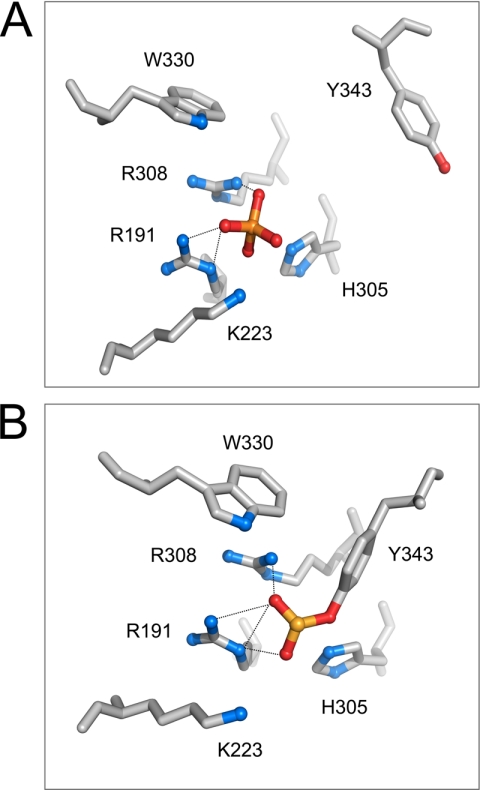
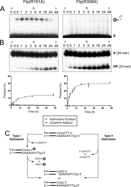
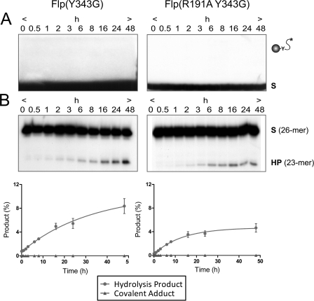
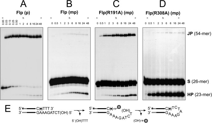
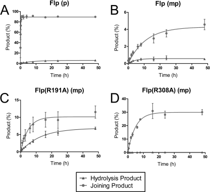
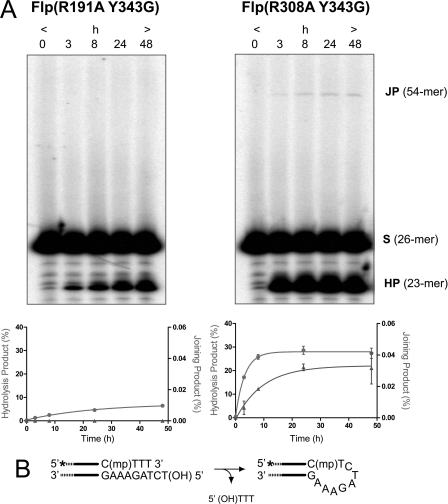
The active site of the tyrosine family site-specific recombinase Flp contains a conserved catalytic pentad that includes two arginine residues, Arg-191 and Arg-308. Both arginines are essential for the transesterification steps of strand cleavage and strand joining in DNA substrates containing a phosphate group at the scissile position. During strand cleavage, the active site tyrosine supplies the nucleophile to form a covalent 3'-phosphotyrosyl intermediate. The 5'-hydroxyl group produced by cleavage provides the nucleophile to re-form a 3'-5' phosphodiester bond in a recombinant DNA strand. In previous work we showed that substitution of the scissile phosphate (P) by the charge neutral methylphosphonate (MeP) makes Arg-308 dispensable during the catalytic activation of the MeP diester bond. However, in the Flp(R308A) reaction, water out-competes the tyrosine nucleophile (Tyr-343) to cause direct hydrolysis of the MeP diester bond. We now report that for MeP activation Arg-191 is also not required. In contrast to Flp(R308A), Flp(R191A) primarily mediates normal cleavage by Tyr-343 but also exhibits a weaker direct hydrolytic activity. The cleaved MeP-tyrosyl intermediate formed by Flp(R191A) can be targeted for nucleophilic attack by a 5'-hydroxyl or water and channeled toward strand joining or hydrolysis, respectively. In collaboration with wild type Flp, Flp(R191A) promotes strand exchange between MeP- and P-DNA partners. Loss of a catalytically crucial positively charged side chain can thus be suppressed by a compensatory modification in the DNA substrate that neutralizes the negative charge on the scissile phosphate.


Figures

FIGURE 1.
FIGURE 1.
Organization of active site residues in DNA-bound Flp with the scissile phosphate in its uncleaved and cleaved (covalently linked) states. The catalytic pentad residues (Arg-191, Lys-K223, His-305, Arg-308, and Trp-330) of a Flp monomer and the active site tyrosine (Tyr-343) donated in trans from a second monomer are displayed surrounding the scissile phosphate (gold sphere). In one active site configuration (A), the scissile phosphate is not cleaved; in the other (B), the scissile phosphate is cleaved and linked to the trans-donated Tyr-343. Note that Arg-191 and Arg-308 make hydrogen bonding interactions with the nonbridging oxygens of the scissile phosphate in both A and B (dotted lines). The structural information is from Protein Data Bank entry 1M6X (21) and is displayed using the PyMOL molecular graphics system (DeLano Scientific).
FIGURE 2.
FIGURE 2.
Endonuclease activities of Flp(R191A) and Flp(R308A) on a MeP half-site substrate. A and B, the reactions were analyzed simultaneously by SDS-PAGE (A) for formation of covalent Flp-DNA adduct by Tyr-343-mediated strand cleavage and by denaturing urea-PAGE (B) for formation of the 23-mer hydrolysis product (HP). The end-labeled double-stranded DNA half-site substrate (A) or the labeled single-stranded DNA (26-mer) resulting from its denaturation (B) is denoted by (S). The 5′-OH on the bottom strand of the substrate was phosphorylated to prevent “strand joining.” The respective plots for Flp(R191A) and Flp(R308A) represent the mean values from three independent experiments with the standard errors indicated by bars. C, type I and type II endonuclease activities leading to hydrolysis of the MeP-tyrosyl intermediate and direct hydrolysis of MeP, respectively, are schematically depicted. The asterisk indicates a 32P-label at the 5′-end of the scissile top strand; (mp), (p), and (HO) represent the scissile MeP, 5′-terminal phosphate, and 5′-terminal hydroxyl, respectively. The filled circle represents Flp recombinase with its active site tyrosine (Y).
FIGURE 3.
FIGURE 3.
Type II endonuclease activities of Flp(Y343G) and Flp(R191A,Y343G) on the MeP half-site. The reactions were performed and analyzed as described for Fig. 2. The results from three separate experiments are plotted.
FIGURE 4.
FIGURE 4.
Strand joining by Flp, Flp(R191A), and Flp(R308A) assayed using P half-site and MeP half-site substrates. The half-site substrates in the strand joining assays contained a free 5′-hydroxyl on the bottom (nonscissile) strand. A–D, reactions were analyzed by denaturing urea-PAGE to detect the products of strand joining (JP) and hydrolysis (HP). A and B, the reactions of wild type Flp with the P half-site (A) or the MeP half-site (B) are shown. C and D, only the MeP half-site reactions are shown for Flp(R191A) (C) and Flp(R308A) (D) because they were inactive on the P half-site (data not shown). The strand joining reaction mediated by the attack of the bottom strand 5′-OH on the P- or MeP-tyrosyl intermediate is schematically illustrated in E. The asterisk indicates the 32P label at the 5′-end of the top (scissile) strand, (●) denotes the scissile P or MeP, and (HO) represents the 5′-terminal hydroxyl group.
FIGURE 5.
FIGURE 5.
Quantitative analysis of the strand joining and endonuclease activities of Flp, Flp(R191A), and Flp(R308A) on half-site substrates containing a 5′-OH on the nonscissile strand. The kinetics of strand joining and hydrolysis are represented by the blue and red circles, respectively.
FIGURE 6.
FIGURE 6.
Strand joining activity of Flp(R308A,Y343G) in the MeP half-site substrate by potential direct attack of the DNA backbone by the 5′-OH. A, joining reactions with Flp(R191A,Y343G) and Flp(R308A,Y343G) were performed and analyzed as described for Fig. 4. The plots below represent three independent experiments. B, schematic diagram for a potential strand joining reaction.
FIGURE 7.
FIGURE 7.
Strand exchange between a P full-site and a MeP half-site promoted collaboratively by Flp and Flp(R191A). A, the exchange reactions between the labeled MeP half-site and the unlabeled P full-site are schematically represented, with the thin wavy line representing the ester bond formed between MeP and the 5′-hydroxyl group. B, the predicted lengths of the labeled recombinant strands resulting from the two possible orientations of the half-site with respect to the full-site are 186 and 130 nucleotides, respectively. The reactions were carried out with a fixed amount of Flp and increasing amounts of Flp(R191A). The reactions were analyzed by 6% urea-PAGE gels. S(26-mer) stands for the labeled strand of the MeP half-site, and RP1(186-mer) and RP2(131-mer) stand for the labeled strands in the recombinant products.

Similar articles

Cited by

References

    1. Perry K., Hwang Y., Bushman F. D., Van Duyne G. D. (2006) Mol. Cell 23, 343–354 - PubMed
    1. Grindley N. D., Whiteson K. L., Rice P. A. (2006) Annu. Rev. Biochem. 75, 567–605 - PubMed
    1. Chen Y., Rice P. A. (2003) Annu. Rev. Biophys. Biomol. Struct. 32, 135–159 - PubMed
    1. Grainge I., Jayaram M. (1999) Mol. Microbiol. 33, 449–456 - PubMed
    1. Guo F., Gopaul D. N., van Duyne G. D. (1997) Nature 389, 40–46 - PubMed

Publication types

LinkOut - more resources