. 2010 Jul 9;285(28):21590-9.
doi: 10.1074/jbc.M109.070169. Epub 2010 May 6.

Peroxisome proliferator-activated receptor gamma co-activator 1alpha (PGC-1alpha) and sirtuin 1 (SIRT1) reside in mitochondria: possible direct function in mitochondrial biogenesis

Affiliations

Peroxisome proliferator-activated receptor gamma co-activator 1alpha (PGC-1alpha) and sirtuin 1 (SIRT1) reside in mitochondria: possible direct function in mitochondrial biogenesis

Katia Aquilano et al. J Biol Chem. .

Abstract

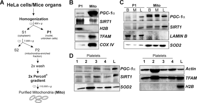
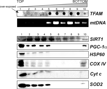
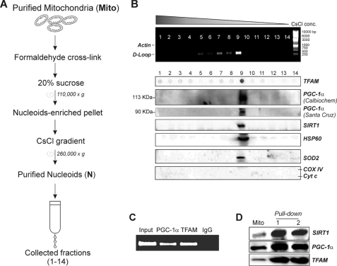
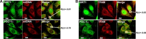
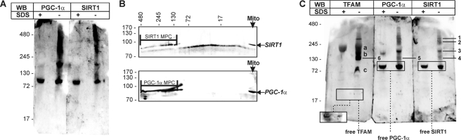
The transcriptional co-activator PGC-1alpha and the NAD(+)-dependent deacetylase SIRT1 are considered important inducers of mitochondrial biogenesis because in the nucleus they regulate transcription of nucleus-encoded mitochondrial genes. We demonstrate that PGC-1alpha and SIRT1 are also present inside mitochondria and are in close proximity to mtDNA. They interact with mitochondrial transcription factor A (TFAM) as assessed by confocal microscopy analysis and by blue native-PAGE. Nucleoid purification allowed us to identify SIRT1 and PGC-1alpha as proteins associated with native and cross-linked nucleoids, respectively. After mtDNA immunoprecipitation analysis, carried out on mitochondrial extracts, we found that PGC-1alpha is present on the same D-loop region recognized by TFAM. Finally, by oligonucleotide pulldown assay, we found PGC-1alpha and SIRT1 associated with the TFAM consensus sequence (human mitochondrial transcription factor-binding site H). The results obtained suggest that in mitochondria PGC-1alpha and SIRT1 may function as their nuclear counterparts and represent the genuine factors mediating the cross-talk between nuclear and mitochondrial genome. Finally, this work adds new knowledge on the function of SIRT1 and PGC-1alpha and highlights the direct involvement of such proteins in regulation of mitochondrial biogenesis.


Figures

FIGURE 1.
FIGURE 1.
Detection of mitochondrial SIRT1 and PGC-1α by immunofluorescence microscopy in SH-SY5Y cells. A, mitochondrial network and nuclei were evidenced by incubation with mouse anti-cytochrome c and Hoechst 33342, respectively. PGC-1α or SIRT1 was stained by using rabbit antibodies (Santa Cruz Biotechnology). B, PGC-1α and SIRT1 were stained by using goat anti-PGC-1α (Santa Cruz Biotechnology) and rabbit anti-SIRT1. Cells were analyzed by an Olympus Delta vision deconvolution fluorescent microscope. The yellow-orange color obtained by image merge indicates the regions where the green and red signals superimpose. White arrows indicate some regions of co-localization signals. Scale bar, 10 μm.
FIGURE 2.
FIGURE 2.
Detection of mitochondrial SIRT1 and PGC-1α in mitochondria purified from HeLa cells and mouse organs and in human platelets. A, scheme for purification of mitochondria from HeLa and mouse organs. B, 10 μg of proteins obtained from HeLa purified mitochondria (Mito) and nuclei-enriched pellets (P1) were subjected to SDS-PAGE followed by Western blot analysis. The possible presence of nuclear protein contaminants was measured by incubating nitrocellulose membrane with rabbit anti-H2B. The effective mitochondria isolation was assessed by staining with rabbit anti-TFAM and mouse anti-cytochrome c oxidase sub IV (COX IV). PGC-1α and SIRT1 were detected by using rabbit Santa Cruz Biotechnology antibodies. C, 10 μg of proteins obtained from nuclei-enriched pellets (P1) and mitochondria (Mito) isolated from mice brain (B), skeletal muscle (M), and liver (L) were subjected to SDS-PAGE followed by Western blot analysis. The possible presence of nuclear protein contaminants was measured by incubating nitrocellulose membrane with rabbit anti-lamin B. The effective mitochondrial isolation was assessed by staining with rabbit anti-SOD2. PGC-1α and SIRT1 were detected by using Santa Cruz Biotechnology antibodies. D, human platelets were purified from peripheral blood, and 20 μg of protein extracts were subjected to SDS-PAGE followed by Western blot analysis. The possible presence of nuclear proteins was measured by incubating the nitrocellulose membrane with rabbit anti-H2B. PGC-1α and SIRT1 were detected by using Santa Cruz Biotechnology antibodies. Immunoblots reported are from one experiment representative of at least three that gave similar results.
FIGURE 3.
FIGURE 3.
Assay of the presence of SIRT1 and PGC-1α in mitochondrial native nucleoids. Native nucleoids were obtained from isolated liver mitochondria as described under “Experimental Procedures.” After sucrose gradient, nucleoid-containing fractions were identified by detecting the presence of TFAM by dot blot (using goat anti-TFAM) and mtDNA by PCR analysis of the D-loop region. Western blot analyses with mouse anti-cytochrome c oxidase subunit IV (COX IV), mouse anti-cytochrome c (Cyt c), rabbit anti-SOD2, and rabbit anti-HSP60 were carried out to determine the possible presence of protein contaminants. Fractions 6–9 were considered to contain native nucleoids due to the absence of protein contaminants and to the presence of TFAM and mtDNA. PGC-1α and SIRT1 were detected by using Santa Cruz Biotechnology antibodies. Immunoblots reported are from one experiment representative of at least three that gave similar results.
FIGURE 4.
FIGURE 4.
Assay of the presence of SIRT1 and PGC-1α in cross-linked mitochondrial nucleoids. A, scheme for purification of nucleoids (N) from mouse liver mitochondria. Fractions (1–14) were finally collected by pricking the bottom of the tube. B, presence of mtDNA in the collected fractions was assayed performing PCR analysis of D-loop region. The presence of genomic DNA was excluded by performing PCR analysis of actin gene. After cross-linking reversion, fractions were subjected to SDS-PAGE. Western blot analysis with mouse anti-cytochrome c oxidase subunit IV (COX IV) and mouse anti-cytochrome c (Cyt c) was carried out to determine the possible presence of protein contaminants. SIRT1, HSP60, TFAM, and SOD2 were detected by using specific rabbit antibodies. PGC-1α was detected by using both the Santa Cruz Biotechnology and the Calbiochem antibodies. C, cross-linked mitochondria from mouse liver were sonicated and immunoprecipitated with goat anti-TFAM or goat anti-PGC-1α antibody (Santa Cruz Biotechnology). DNA was extracted, and PCR analysis of the region +15,600/+15,868 of mtDNA was successively carried out. Input and immunoprecipitation with a control IgG were used as positive and negative controls. D, mitochondrial protein extracts from HEK293 cells were analyzed by oligonucleotide pulldown assay using TFAM biotinylated consensus oligonucleotide and Western blot analysis using Santa Cruz Biotechnology anti-TFAM, anti-SIRT1, and anti-PGC-1α antibody. Mito, total mitochondrial lysates; lanes 1 and 2, two independent oligonucleotide pulldown experiments. Immunoblots reported are from one experiment representative of at least three that gave similar results.
FIGURE 5.
FIGURE 5.
Determination of the association of SIRT1 and PGC-1α with mtDNA and TFAM by confocal microscopy in HeLa cells. A, mtDNA was labeled by BrdUrd incorporation followed by methanol-acetone fixing and immunostaining with mouse anti-BrdUrd. SIRT1 or PGC-1α was also immunostained using Santa Cruz Biotechnology antibodies. Cells were then visualized by confocal microscopy, and the level of superimposition of SIRT1 or PGC-1α-derived signals with that of BrdUrd was evaluated by calculation of the Pearson's correlation coefficient (R(r)). Values >0.50 were considered to be significant. B, HeLa cells were fixed with paraformaldehyde and immunostained with goat anti-TFAM and rabbit Santa Cruz Biotechnology anti-PGC-1α or SIRT1. Cells were then visualized by confocal microscopy, and the level of superimposition of SIRT1 or PGC-1α-derived signals with that of TFAM was evaluated by calculation of the Pearson's correlation coefficient (R(r)). Values >0.50 were considered to be significant. Scale bar, 10 μm.
FIGURE 6.
FIGURE 6.
Identification of mitochondrial multiprotein complexes by BN-PAGE in liver purified mitochondria. A, mitochondrial proteins were extracted from liver purified mitochondria (mito) using a lysis buffer maintaining native interaction in multiprotein complexes. Ten μg of proteins were loaded on blue native gel. The same samples were boiled in the presence of SDS and loaded in parallel with the respective native sample. Western blot (WB) analysis using rabbit Santa Cruz Biotechnology anti-SIRT1 or anti-PGC-1α was then performed. B, after BN-PAGE, a second SDS-PAGE dimension was carried out followed by Western blot analysis using rabbit Santa Cruz Biotechnology anti-PGC-1α or anti-SIRT1. MPC, multiprotein complexes. C, 10 μg of proteins were loaded on blue native gel, and Western blot using a rabbit anti-TFAM was carried out. 1, multiprotein complex containing TFAM, PGC-1α, and SIRT1; 2 and 3, multiprotein complexes containing PGC-1α and SIRT1; 4, multiprotein complex containing TFAM, and SIRT1; 5 multiprotein complex containing TFAM and PGC-1α; 6, multiprotein complex containing TFAM-PGC-1α and TFAM-SIRT1; a–c, other TFAM multiprotein complexes. To visualize free TFAM protein, a major exposure was mandatory due to the lower cross-reactivity of TFAM antibody against native TFAM with respect to TFAM engaged in multiprotein complexes (see inset in TFAM immunoblot). Immunoblots reported are from one experiment representative of four that gave similar results.

Similar articles

Cited by

References

    1. Hock M. B., Kralli A. (2009) Annu. Rev. Physiol. 71, 177–203 - PubMed
    1. Chow L. S., Greenlund L. J., Asmann Y. W., Short K. R., McCrady S. K., Levine J. A., Nair K. S. (2007) J. Appl. Physiol. 102, 1078–1089 - PubMed
    1. Scarpulla R. C. (2008) Physiol. Rev. 88, 611–638 - PubMed
    1. Guarente L. (2007) Cold Spring Harbor Symp. Quant. Biol. 72, 483–488 - PubMed
    1. Rodgers J. T., Lerin C., Gerhart-Hines Z., Puigserver P. (2008) FEBS Lett. 582, 46–53 - PMC - PubMed

Publication types

LinkOut - more resources