. 2011 Mar;44(3):350-60.
doi: 10.1165/rcmb.2009-0422OC. Epub 2010 May 6.

Neutral sphingomyelinase 2: a novel target in cigarette smoke-induced apoptosis and lung injury

Affiliations

Neutral sphingomyelinase 2: a novel target in cigarette smoke-induced apoptosis and lung injury

Simone Filosto et al. Am J Respir Cell Mol Biol. 2011 Mar.

Abstract

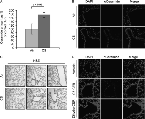
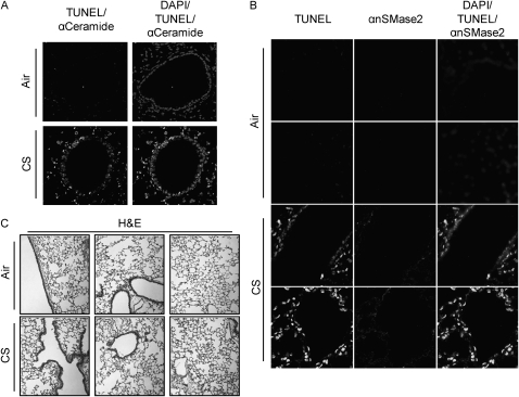
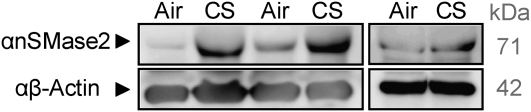
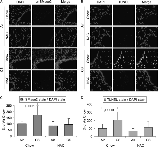
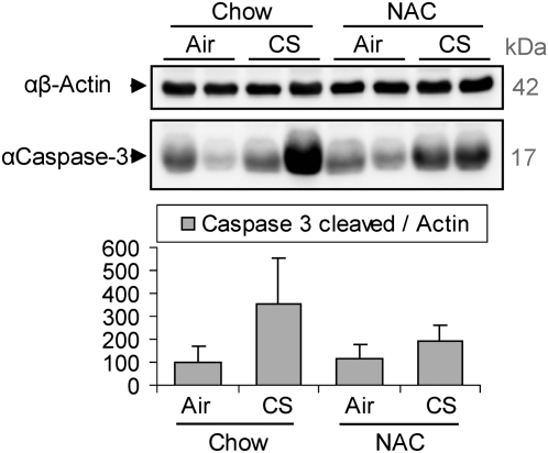
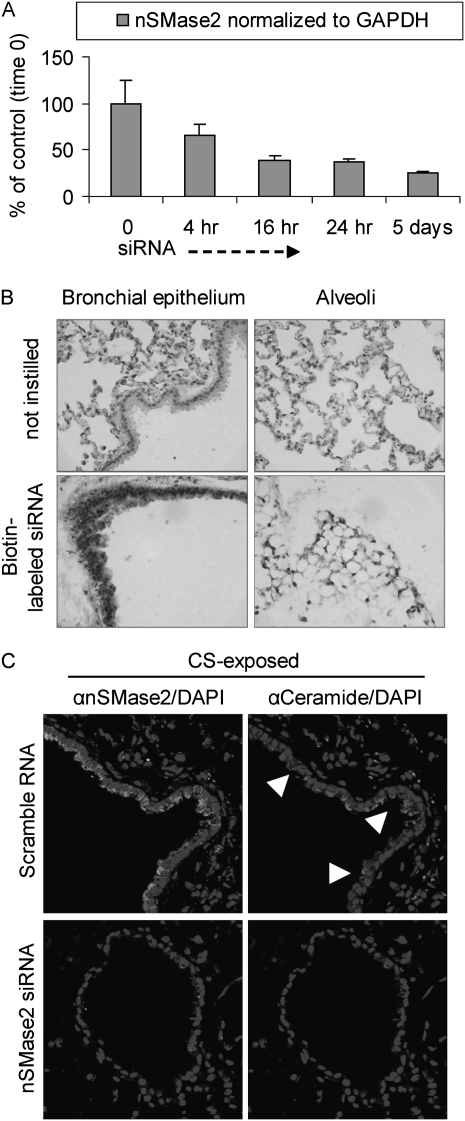
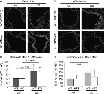
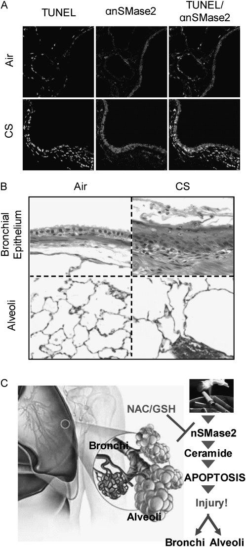
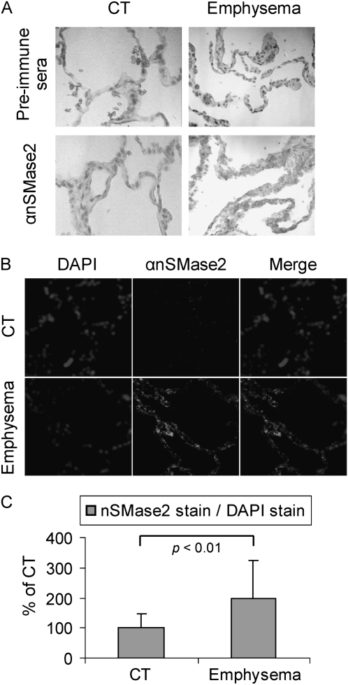
Chronic obstructive pulmonary disease (COPD) is caused by exposure to cigarette smoke (CS). One mechanism of CS-induced lung injury is aberrant generation of ceramide, which leads to elevated apoptosis of epithelial and endothelial cells in the alveolar spaces. Recently, we discovered that CS-induced ceramide generation and apoptosis in pulmonary cells is governed by neutral sphingomyelinase (nSMase) 2. In the current experiments, we expanded our studies to investigate whether nSMase2 governs ceramide generation and apoptosis in vivo using rodent and human models of CS-induced lung injury. We found that exposure of mice or rats to CS leads to colocalizing elevations of ceramide levels and terminal deoxynucleotidyl transferase mediated X-dUTP nick end labeling-positive cells in lung tissues. These increases are nSMase2 dependent, and are abrogated by treatment with N-acetyl cysteine or anti-nSMase2 small interfering RNA (siRNA). We further showed that mice that are heterozygous for nSMase2 demonstrate significant decrease in ceramide generation after CS exposure, whereas acidic sphingomyelinase (aSMase) knockout mice maintain wild-type ceramide levels, confirming our previous findings (in human airway epithelial cells) that only nSMase2, and not aSMase, is activated by CS exposure. Lastly, we found that lung tissues from patients with emphysema (smokers) display significantly higher levels of nSMase2 expression compared with lung tissues from healthy control subjects. Taken together, these data establish the central in vivo role of nSMase2 in ceramide generation, aberrant apoptosis, and lung injury under CS exposure, underscoring its promise as a novel target for the prevention of CS-induced airspace destruction.


Figures

Figure 1.
Figure 1.
Cigarette smoke (CS) induces ceramide generation in the lung. Three 129/Sv mice for each time/treatment point were exposed, or not, to CS for 1 week (A and B), whereas two 129/Sv mice for each treatment/point were intratracheally instilled with ceramide analogs or BSA (vehicle) (D). The mice were killed and assayed for ceramide levels in the lung by diacylglycerol kinase assay (A) and immunohistochemistry (IHC) (B and D). (A) The amount of ceramide in the graphic represents the average of three independent experiments (three mice per treatment), and is reported as percentage of the untreated mice (filtered air) after normalization per protein unit of the tissue; the P value in (A) was obtained by Student's t test per number of mice. (B) Total nuclei (left panels) were stained by 4′,6-diamidino-2-phenylindole (DAPI ceramide (central panels) was localized by incubating lung slides with a specific anti- (α) ceramide antibody (Ab) and stained by Alexa Fluor 555 dye–conjugated Ab; the panels on the right show the merges of total nuclei and ceramide staining. (C) Hematoxylin and eosin (H&E) stain of the 129/Sv mice exposed, or not, to CS for 1 week. (D) mice were instilled with synthetic lipids, either C6-ceramide (C6-CER) or dihydro-C6-ceramide (Dihydro-CER), killed 24 hours after instillation, and assayed for ceramide levels in the lung by IHC: ceramide (central panels) was localized as in (A) and stained by Alexa Fluor 555 dye; total nuclei were stained by DAPI (left panels); merge images of ceramide and nuclei staining are shown (right panels). Images were acquired with an LSM 5 Pascal Zeiss laser scanning or an Olympus FluoView FV1000 confocal microscope.
Figure 2.
Figure 2.
Ceramide generation, neutral sphingomyelinase (nSMase) 2 overexpression, and DNA fragmentation are strictly related in lungs of CS-exposed mice. (A) C57BL/6 mice were exposed, or not, to CS for 4 weeks, then killed and assayed for ceramide levels by IHC, as in Figure 1 and fragmented DNA by terminal deoxynucleotidyl transferasemediated X-dUTP nick end labeling (TUNEL) assay (brighter stain); total nuclei were also stained by DAPI, as in Figure 1. The left panel shows the merge between fragmented DNA and ceramide staining, whereas the right panel presents the merge of total nuclei, fragmented DNA (brighter stain), and ceramide localization. (B) Mice were exposed, or not, to CS for 5 weeks, killed, and tested for fragmented DNA as in (A) (stain in left panels) and nSMase2 expression (stain in second panels from the left) by incubating lung slides with a specific anti- (α) nSMase2 Ab, and staining it by Alexa Fluor 555 dye. Total nuclei were stained by DAPI (not shown alone); right panels show the merge of TUNEL (brighter stain), αnSMase2, and DAPI. (C) H&E stain (overall lung histology) of the C57BL/6 mice exposed, or not, to CS for 5 weeks.
Figure 3.
Figure 3.
nSMase2 is overexpressed in 129/Sv mice after 1 week of exposure to CS. Three 129/Sv mice were exposed, or not, to CS for 1 week, then killed, and assayed by immunoblotting (IB) for the levels of nSMase2 expression in the whole-lung lysates (lung tissues homogenized in the presence of 0.4% Triton-X 100). Total protein (200 μg) from each lung was separated on SDS-PAGE and immunoblotted using specific αnSMase2 and αβ-actin antibodies. Each sample in the figure represents a different mouse. Separate panels represent two different Western blot analyses.
Figure 4.
Figure 4.
N-acetyl cysteine (NAC) counteracts nSMase2 overexpression and DNA fragmentation induced by CS. Three C57BL/6 mice were exposed, or not, to CS for 4 weeks, and fed, or not, with a 0.4% NAC–enriched diet, then killed, and assayed by IHC for nSMase2 expression (A) or by TUNEL staining for detection of fragmented DNA (B). Total nuclei were stained by DAPI (left panels); nSMase2 was stained by Alexa Fluor 555 dye (central panels in [A]); TUNEL-positive cells are in central panels in [B]; respective merge images are shown in the right panels. (C) A total of 12 random images were acquired at 200× magnification from lung sections of three different mice per each treatment (four images per each mouse) and the mean intensity of nSMase2 stain (analyzed by LSM 5 Pascal 4.2 software) was normalized to the mean intensity of DAPI stain (proportional to cell nuclei/number) in each image; then, the average value of nSMase2 stain from the air/chow–treated mice was arbitrarily set as 100% value, and all other values (means and SD) have been adjusted accordingly. (D) TUNEL stain in the lung sections was quantified as described above in (C) and reported as means (±SDs). The P values in (C) and (D) were obtained by Student's t test per number of images.
Figure 5.
Figure 5.
CS induces caspase-3 activation, which is reduced by NAC. Three C57BL/6 mice were exposed, or not, to CS for 5 weeks, and fed, or not, with a 0.4% NAC enriched diet, then killed, and assayed by IB for the activation of caspase-3 (presence of cleaved caspase-3) in the whole-lung homogenates. The image shows samples of 200 μg total proteins from lungs of two different mice per treatment. The samples were separated on SDS-PAGE and immunoblotted using specific αβ-actin and αcaspase-3 antibodies. The histogram below shows the quantification (by densitometry) of the levels of caspase-3 cleaved normalized to the respective levels of β-actin in the lungs of three different mice.
Figure 6.
Figure 6.
Small interfering RNA (siRNA) of nSMase2 in the lung prevents ceramide accumulation during CS exposure. Two 129/Sv mice for each time/treatment point were instilled for up to 5 days with biotin-labeled nSMase2 siRNA or scrambled RNA, while exposed in parallel to CS. (A) nSMase2 expression in siRNA-silenced lungs was evaluated by RT-PCR in triplicate samples and normalized to the level of glyceraldehyde 3-phosphate dehydrogenase (GAPDH). (B) Lung sections from 5-day-instilled mice were incubated with peroxidase-labeled streptavidin and 3,3′-diaminobenzidine (DAB) substrate to assess the presence of biotin (darker staining), which indicates the presence of siRNA both in airway and in alveoli (left and right panels, respectively); images were acquired by optical microscopy. (C) Lung sections from mice instilled with nSMase2 siRNA for 5 days, or instilled with scrambled RNA, were subjected to IHC using specific αnSMase2 (brighter stain in left panels) and αceramide (brighter stain in right panels) antibodies; total nuclei were stained by DAPI; arrowheads indicate examples of ceramide accumulation–positive cells. Fluorescent images were acquired with an Olympus FluoView FV1000 laser scanning confocal microscope and show the respective merges of nSMase2/nuclei and ceramide/nuclei stains in mice instilled with either scrambled siRNA (upper panels) or nSMase2 siRNA (lower panels).
Figure 7.
Figure 7.
nSMase2, and not acidic sphingomyelinase (aSMase), is responsible for ceramide generation during CS exposure. Knockout (KO) mice for aSMase (A) and heterozygous (HET) mice for nSMase2 (nSMase2−/+) (B) were exposed, or not, to CS for 3 weeks, killed, and assayed by IHC for ceramide levels in the lung sections using α-ceramide Ab, as in Figure 1 (brighter stain). Total nuclei were stained by DAPI. Images were acquired with an Olympus FluoView FV1000 laser scanning confocal microscope and show the merges of respective total nuclei and ceramide stains in filtered air (left panels) and CS-exposed (right panels) mice, for both KO aSMase (A) and HET nSMase2 (B), compared with the respective wild-type (WT) control animals (upper panels). (C) A total of 16 random images were acquired at 200× magnification from lung sections of 2 different mice per each treatment (8 images per each mouse), and the mean intensity of ceramide stain has been normalized to DAPI stain as in Figure 4 (i.e., the average values of ceramide stains from the air-exposed mice), for both WT and KO aSMase mice, were arbitrarily set as 100%, and other values (means and SDs) were adjusted. (D) Ceramide stains in lung sections of 129/Sv mice, WT, or HET nSMase2, were quantified as described in (C) and reported as means (±SD). The P values in (C) and (D) were obtained by Student's t test per number of images.
Figure 8.
Figure 8.
nSMase2 expression and TUNEL staining are augmented in lung of CS-exposed rats. Spontaneously hypertensive rat (SHR)/NCrIBR rats were exposed, or not, to CS. (A) Fragmented DNA was stained by TUNEL assay as in Figure 2 (left panels), and nSMase2 expression was evaluated by incubation with αnSMase2 Ab (central panels). Merge images of TUNEL and nSMase2 staining are shown in the right panels. Images were acquired with an Olympus FluoView FV1000 laser scanning confocal microscope. (A) Representative of images scored by four people independently, who analyzed random lung sections. (B) Lung sections stained by H&E showed epithelial metaplasia (upper panels) and parenchyma airspace enlargement (lower panels) after CS exposure: the epithelial lining of the airway in the filtered air control is composed of a simple cuboidal layer of ciliated and nonciliated cells (upper left panel). In contrast, exposure to tobacco smoke for 14 weeks is associated with a thickened, stratified squamous epithelium in the form of keratinizing squamation of the epithelium, with numerous inflammatory cells and debris in the airway lumen (upper right panel). The parenchyma (lower panels) show a significant enlargement of the airspaces of CS-exposed mice (lower right panel) in comparison to the filtered air-exposed control (lower left panel). The changes in airspace size are estimated to be increased approximately 50% above filtered air controls in mean linear intercept length within the airspaces of the lung parenchyma. (C) Model of CS-induced injury. CS exposure of airway cells specifically causes overexpression and activation of nSMase2, which leads to increased ceramide generation and occurrence of apoptosis, producing lung injury in both bronchial epithelium and alveoli. An antioxidant, such as NAC, precursor of glutathione (GSH), can quench the overexpression and activation of nSMase2, and thus reduce the occurrence of apoptosis.
Figure 9.
Figure 9.
nSMase2 expression is augmented in lung tissue from patients with emphysema. Lung tissue sections were incubated with αnSMase2 Ab or the preimmune sera (rabbit). Images were acquired by optical microscopy after colorimetric staining by incubating with avidin–biotin complex Ab and DAB (A), or with an LSM 5 Pascal Zeiss laser scanning confocal microscope after staining αnSMase2 by Alexa Fluor 555 dye (central panels) (B). (A) Representative of images scored by four people independently, who analyzed random lung sections from control subjects (CTs) or patients with emphysema. (B) Total nuclei were stained by DAPI and merged with images of nSMase2 expression. The histogram in (C) shows the quantification of nSMase2 staining in the fluorescent experiments. A total of 16 random images was acquired at 200× magnification from lung sections of 2 different CTs and patients with emphysema (8 images per patient), then the mean intensity of nSMase2 stain was normalized to DAPI stain, as in Figure 4. The average value of nSMase2 stains of the CTs was arbitrarily set as 100%, and the other values (means and SDs) were adjusted. The P value was obtained by Student's t test per number of images.

Similar articles

Cited by

References

    1. Yoshida T, Tuder RM. Pathobiology of cigarette smoke–induced chronic obstructive pulmonary disease. Physiol Rev 2007;87:1047–1082. - PubMed
    1. Churg A, Wright JL. Proteases and emphysema. Curr Opin Pulm Med 2005;11:153–159. - PubMed
    1. Demedts IK, Demoor T, Bracke KR, Joos GF, Brusselle GG. Role of apoptosis in the pathogenesis of COPD and pulmonary emphysema. Respir Res 2006;7:53. - PMC - PubMed
    1. Giordano RJ, Lahdenranta J, Zhen L, Chukwueke U, Petrache I, Langley RR, Fidler IJ, Pasqualini R, Tuder RM, Arap W. Targeted induction of lung endothelial cell apoptosis causes emphysema-like changes in the mouse. J Biol Chem 2008;283:29447–29460. - PMC - PubMed
    1. Petrache I, Natarajan V, Zhen L, Medler TR, Richter AT, Cho C, Hubbard WC, Berdyshev EV, Tuder RM. Ceramide upregulation causes pulmonary cell apoptosis and emphysema-like disease in mice. Nat Med 2005;11:491–498. - PMC - PubMed

Publication types