Comparative Study
. 2010 Jul 9;107(1):106-16.
doi: 10.1161/CIRCRESAHA.109.214601. Epub 2010 May 6.

Therapeutic targeting of mitochondrial superoxide in hypertension

Affiliations
Comparative Study

Therapeutic targeting of mitochondrial superoxide in hypertension

Anna E Dikalova et al. Circ Res. .

Abstract

Rationale: Superoxide (O2(-) ) has been implicated in the pathogenesis of many human diseases including hypertension; however, commonly used antioxidants have proven ineffective in clinical trials. It is possible that these agents are not adequately delivered to the subcellular sites of superoxide production.

Objective: Because the mitochondria are important sources of reactive oxygen species, we postulated that mitochondrial targeting of superoxide scavenging would have therapeutic benefit.

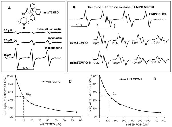
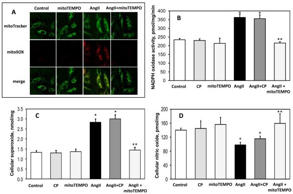
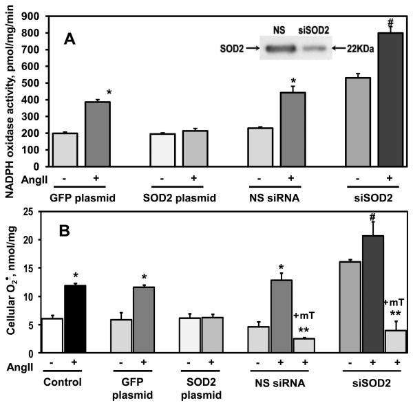
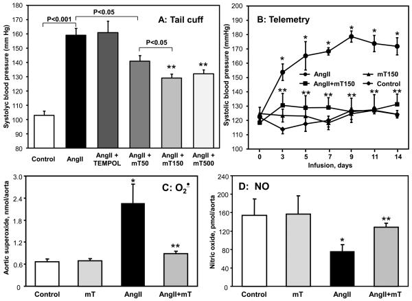
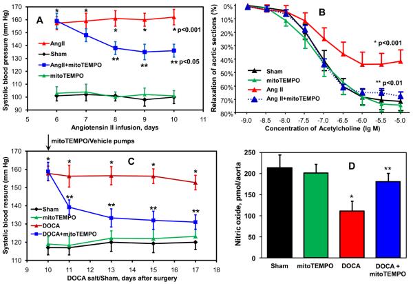
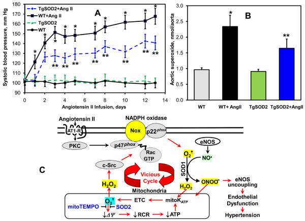
Methods and results: In this study, we found that the hormone angiotensin (Ang II) increased endothelial mitochondrial superoxide production. Treatment with the mitochondria-targeted antioxidant mitoTEMPO decreased mitochondrial O2(-), inhibited the total cellular O2(-), reduced cellular NADPH oxidase activity, and restored the level of bioavailable NO. These effects were mimicked by overexpressing the mitochondrial MnSOD (SOD2), whereas SOD2 depletion with small interfering RNA increased both basal and Ang II-stimulated cellular O2(-). Treatment of mice in vivo with mitoTEMPO attenuated hypertension when given at the onset of Ang II infusion and decreased blood pressure by 30 mm Hg following establishment of both Ang II-induced and DOCA salt hypertension, whereas a similar dose of nontargeted TEMPOL was not effective. In vivo, mitoTEMPO decreased vascular O2(-), increased vascular NO production and improved endothelial-dependent relaxation. Interestingly, transgenic mice overexpressing mitochondrial SOD2 demonstrated attenuated Ang II-induced hypertension and vascular oxidative stress similar to mice treated with mitoTEMPO.

Conclusions: These studies show that mitochondrial O2(-) is important for the development of hypertension and that antioxidant strategies specifically targeting this organelle could have therapeutic benefit in this and possibly other diseases.


Figures

Figure 1
Figure 1
Accumulation of mitoTEMPO in mitochondria and scavenging of O2 by mitoTEMPO. (A) ESR spectra of various compartments from BAEC following incubation with mitoTEMPO (1 μM) for 1 hour. Cells were lysed cytoplasmic and mitochondrial fractions isolated by centrifugation. Spectra of the extracellular media, cellular cytoplasm or mitochondria (10 mg weight for each sample) are shown. (B) Spin trapping of O2 using the spin trap EMPO and xanthine/xanthine oxidase O2 generating system in the absence or presence of mitoTEMPO and mitoTEMPO-H. The efficacy of O2 scavenging was derived from dose-dependent decrease of ESR amplitude of EMPO-OOH in the presence of mitoTEMPO (C) or mitoTEMPO-H (D).
Figure 2
Figure 2
Effect of mitoTEMPO on mitochondrial O2, endothelial O2, nitric oxide and NADPH oxidase activity. (A) Mitochondrial O2 was measured in control or angiotensin II (Ang II)-stimulated HAEC using fluorescent probe MitoSOX. Mitochondrial localization of MitoSOX signal was confirmed by colocalization with MitoTracker. (B) Activity of NADPH oxidase measured in membrane fractions isolated from unstimulated or angiotensin II (Ang II) stimulated BAEC (4 hours, 200nM) and supplemented for 15 minutes with saline, the mitochondria-impermeable SOD mimetic 3-carboxyproxyl (CP), or the mitochondria-targeted SOD mimetic mitoTEMPO (25 nM). (C) Cellular O2 was measured in intact BAEC using DHE and HPLC. (D) Nitric oxide was measured in intact cells after treatment with saline, CP or mitoTEMPO using ESR and the NO spin trap Fe(DETC)2 . Results are mean±SEM, n=5-8 each. *P<0.05 vs control, ** P <0.05 vs Ang II.
Figure 3
Figure 3
SOD2 modulates angiotensin II stimulated O2 production. SOD2 was overexpressed or depleted by 72-hours transfection with a SOD2-expressing plasmid or siRNA. (A) Activity of NADPH oxidase was measured in the membrane fractions of unstimulated or angiotensin II (200 nM, 4 hours) stimulated HAEC using ESR and spin probe CPH . MitoTEMPO (+mT, 25 nM) was added to HAEC after angiotensin II (Ang II) stimulation, 15-minutes prior to isolation of membrane fraction. The insert shows a typical Western blot of mitochondrial fractions isolated from siSOD2 or non-silencing siRNA treated HAEC indicating significant depletion of SOD2 in siSOD2 treated cells. (B) Superoxide was measured by DHE/HPLC . Results are mean±SEM, n=4-8 each, *P<0.01 vs no angiotensin II, **P < 0.01 vs Ang II , §P<0.05 vs NS control, #P < 0.05 vs NS+Ang II .
Figure 4
Figure 4
Attenuation of angiotensin II-induced hypertension in C57Blk/6 mice by mitoTEMPO. (A) Systolic blood pressure in mice infused for 14 days with saline, angiotensin II (Ang II, 0.7 mg/kg/day) or co-infused with mitoTEMPO (50, 150 and 500 μg/kg/day) or TEMPOL. Systolic blood pressure was measured using tail-cuff plethysmography. (B) Telemetric measurements of systolic blood pressure in mice infused with saline, angiotensin II or co-infused with mitoTEMPO (150 μg/kg/day). Blood pressure values represent mean ± SEM for 5-7 animals per group. *P<0.01 vs control, ** P <0.01 vs Ang II. Following 14 days infusion with saline, angiotensin II or co-infusion with mitoTEMPO (150 μg/kg/day) aortic tissue was isolated for measurements of O2 with DHE and HPLC (C) or nitric oxide ESR and Fe(DETC)2 (D). Results represent mean ± SEM for 5-7 animals per group. *P<0.05 vs control, ** P <0.05 vs Ang II.
Figure 5
Figure 5
Effects of mitoTEMPO treatment after the onset of hypertension on blood pressure, vascular function and NO production. (A) Blood pressure of mice treated with mitoTEMPO (0.7 mg/kg/day) after onset of angiotensin II (Ang II)-induced hypertension. (B) Endothelial-dependent relaxation in aortic vessels isolated from mice infused with saline (Sham), mitoTEMPO, angiotensin II or angiotensin II-infused mice treated with mitoTEMPO. Vessels were preconstricted with PGF2α and relaxations to cumulative concentrations of acetylcholine were examined. (C) Blood pressure of mice treated with mitoTEMPO (0.7 mg/kg/day) after onset of DOCA-salt induced hypertension. (D) Production of aortic nitric oxide measured in isolated aorta by ESR and Fe(DETC)2. Results represent mean ± SEM for 6-8 animals per group. *P<0.001 vs Sham, ** P <0.05 vs angiotensin II / DOCA.
Figure 6
Figure 6
Analysis of blood pressure and vascular O2 in SOD2 overexpressing transgenic mice infused with angiotensin II. (A) Systolic blood pressure in TgSOD2 and C57Blk/6 mice infused with saline or angiotensin II (0.7 mg/kg/day). (B) Production of aortic O2 measured with DHE and HPLC. Results represent mean ± SEM for 4-8 animals per group. * P<0.05 vs Ang II. (C) Proposed role of mitochondrial O2 in endothelial dysfunction and hypertension.

Comment in

References

    1. Madamanchi NR, Runge MS. Mitochondrial dysfunction in atherosclerosis. Circ Res. 2007;100:460–473. - PubMed
    1. Puddu P, Puddu GM, Galletti L, Cravero E, Muscari A. Mitochondrial dysfunction as an initiating event in atherogenesis: a plausible hypothesis. Cardiology. 2005;103:137–141. - PubMed
    1. Perier C, Tieu K, Guegan C, Caspersen C, Jackson-Lewis V, Carelli V, Martinuzzi A, Hirano M, Przedborski S, Vila M. Complex I deficiency primes Bax-dependent neuronal apoptosis through mitochondrial oxidative damage. Proc Natl Acad Sci U S A. 2005;102:19126–19131. - PMC - PubMed
    1. Rego AC, Oliveira CR. Mitochondrial dysfunction and reactive oxygen species in excitotoxicity and apoptosis: implications for the pathogenesis of neurodegenerative diseases. Neurochem Res. 2003;28:1563–1574. - PubMed
    1. Wall JA, Wei J, Ly M, Belmont P, Martindale JJ, Tran D, Sun J, Chen WJ, Yu W, Oeller P, Briggs S, Gustafsson AB, Sayen MR, Gottlieb RA, Glembotski CC. Alterations in oxidative phosphorylation complex proteins in the hearts of transgenic mice that overexpress the p38 MAP kinase activator, MAP kinase kinase 6. Am J Physiol Heart Circ Physiol. 2006;291:H2462–72. - PubMed

Publication types