Review
. 2010 May;7(5):277-85.
doi: 10.1038/nrurol.2010.47.

The genetic basis of kidney cancer: a metabolic disease

Affiliations
Review

The genetic basis of kidney cancer: a metabolic disease

W Marston Linehan et al. Nat Rev Urol. 2010 May.

Abstract

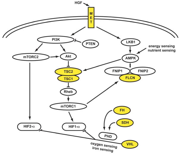
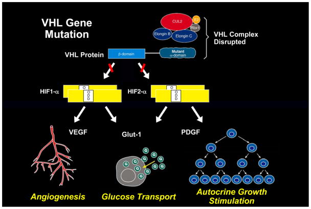
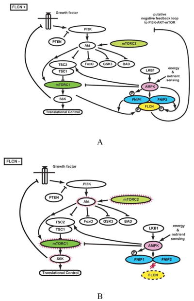
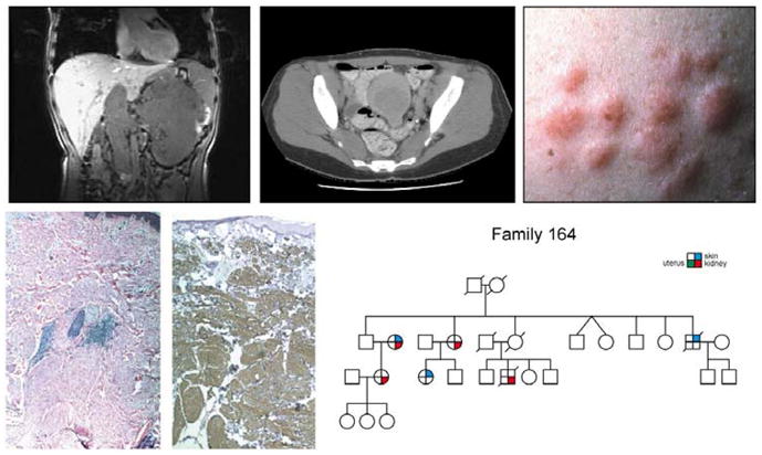
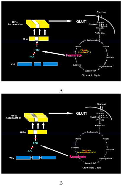
Kidney cancer is not a single disease but comprises a number of different types of cancer that occur in the kidney, each caused by a different gene with a different histology and clinical course that responds differently to therapy. Each of the seven known kidney cancer genes, VHL, MET, FLCN, TSC1, TSC2, FH and SDH, is involved in pathways that respond to metabolic stress or nutrient stimulation. The VHL protein is a component of the oxygen and iron sensing pathway that regulates hypoxia-inducible factor (HIF) levels in the cell. HGF-MET signaling affects the LKB1-AMPK energy sensing cascade. The FLCN-FNIP1-FNIP2 complex binds AMPK and, therefore, might interact with the cellular energy and nutrient sensing pathways AMPK-TSC1/2-mTOR and PI3K-Akt-mTOR. TSC1-TSC2 is downstream of AMPK and negatively regulates mTOR in response to cellular energy deficit. FH and SDH have a central role in the mitochondrial tricarboxylic acid cycle, which is coupled to energy production through oxidative phosphorylation. Mutations in each of these kidney cancer genes result in dysregulation of metabolic pathways involved in oxygen, iron, energy or nutrient sensing, suggesting that kidney cancer is a disease of cell metabolism. Targeting the fundamental metabolic abnormalities in kidney cancer provides a unique opportunity for the development of more-effective forms of therapy for this disease.


Conflict of interest statement

Competing interests

The authors have no competing interests

Figures

Figure 1
Figure 1. The genetic basis of kidney cancer: a metabolic disease
The genes known to cause kidney cancer, VHL, MET, FLCN, FH, SDH, TSC1 and TSC2 share the common feature that each is involved in oxygen, iron, energy and/or nutrient sensing pathways. Kidney cancer is fundamentally a metabolic disease. VHL targets HIF-1α and HIF-2α for ubiquitin-mediated degradation through an oxygen and iron sensing mechanism. The FLCN/FNIP1/FNIP2 complex binds AMPK, the primary energy sensor in the cell, and FLCN is phosphorylated by a rapamycin-sensitive kinase (i.e.,mTORC1). TSC1/TSC2 are phosphorylated by the LKB1/AMPK cascade and help mediate the cell’s response to energy/nutrient sensing. Fumarate hydratase and succinate dehydrogenase are TCA cycle enzymes. When fumarate hydratase or succinate dehydrogenase are deficient, the function of the TCA cycle is impaired and the cell is dependent on glycolysis for energy production. Inactivation of fumarate hydratase or succinate dehydrogenase impairs PHD function and represents a VHL-independent mechanism for dysregulation of HIF degradation. Increased HIF levels lead to increased GLUT1 which enables transport of glucose for ATP production. Abbreviations: folliculin interacting protein 1 (FNIP1) and folliculin interacting protein 2 (FNIP2), HIF prolyl hydroxylase (PHD). Adapted from Linehan, et al.(1)
Figure 2
Figure 2. The VHL complex targets HIF: an oxygen/iron sensing pathway
The VHL E3 ubiquitin ligase complex targets the hypoxia inducible factors for ubiquitin-mediated degradation. This process is mediated by HIF prolyl hydroxylase (PHD) and an essential cofactor, 2-oxoglutarate (2-OG). This is an oxygen/iron sensing system; when the cell is hypoxic or iron levels are low, the VHL complex cannot target and degrade HIF and HIF over-accumulates, driving the transcription of a number of genes important in cancer, such as vascular endothelial growth factor (VEGF), platelet derived growth factor (PDGF) and the glucose transporter, GLUT 1.(–4)
Figure 3
Figure 3. The VHL complex targets HIF-1α and HIF-2α for degradation
The VHL complex targets HIF-1α and HIF-2α for ubiquitin mediated degradation.(2) When the VHL gene is mutated, as in clear cell kidney cancer, the complex cannot target and degrade HIF. HIF-2α is thought to be a critical pathway for VHL-deficient clear cell kidney tumorigenesis.(5;6)
Figure 4
Figure 4. Birt-Hogg-Dubé (BHD) syndrome: kidney cancer, cutaneous fibrofolliculomas and pulmonary cysts
BHD patients are at risk for the development of benign cutaneous tumors (A, B) that may occur on the face and neck. These cutaneous lesions are benign fibrofolliculomas (C). BHD patients are at risk for the development of renal tumors that can be solitary (D), bilateral, multifocal (E) and can grow to large size and metastasize (F). From Pavlovich, et al.(7)
Figure 5
Figure 5. The FLCN pathway
A. FLCN is the gene for the Birt-Hogg-Dubé (BHD) syndrome. Patients affected with BHD are characterized by germline mutation of the FLCN gene. The FLCN/FNIP1/FNIP2 complex binds AMPK and FLCN is phosphorylated by a rapamycin-sensitive kinase (i.e.,mTORC1). B. When FLCN is deficient, AKT, mTORC1 and mTORC2 are activated. From Hasumi, et al.(8)
Figure 6
Figure 6. Hereditary leiomyomatosis renal cell cancer
Hereditary leiomyomatosis renal cell cancer (HLRCC) is a hereditary cancer syndrome in which affected individuals are at risk for the development of kidney cancer (upper left panel) and uterine (upper middle panel) and cutaneous (upper right panel) leiomyomas (lower left and center panels). From Linehan et al.(9)
Figure 7
Figure 7. Fumarate Hydratase and Succinate Dehydrogenase Deficient Kidney Cancer: the Warburg Effect
Fumarate hydratase and succinate dehydrogenase–deficient kidney cancer are examples of the Warburg effect in cancer.(10) Germline fumarate hydratase mutations are found in the germline of patients affected with Hereditary Leiomyomatosis Renal Cell Cancer (HLRCC). HLRCC patients are at risk for the development of an aggressive form of kidney cancer. A) In fumarate hydratase deficient cells, fumarate accumulates and inhibits prolyl hydroxylation by competitively inhibiting HPH (A). HIF accumulates and increases the transcription of vascular endothelial growth factor and the glucose transporter, GLUT-1. Oxidative phosphorylation is impaired by the loss of the TCA cycle enzyme, fumarate hydratase, and the cells depend on glycolysis for energy production.(11;12) B) Succinate dehydrogenase is another TCA cycle enzyme which has also been associated with familial renal carcinoma. Increased levels of succinate have also been shown to inhibit HPH and a similar mechanism for tumorigenesis is likely for SDH-associated kidney cancer.(11)

References

    1. Linehan WM, Walther MM, Zbar B. The genetic basis of cancer of the kidney. J Urol. 2003 Dec;170(6 Pt 1):2163–72. - PubMed
    1. Vogelstein B, Kinzler KW. Cancer genes and the pathways they control. Nat Med. 2004 Aug;10(8):789–99. - PubMed
    1. Thompson CB. Attacking cancer at its root. Cell. 2009 Sep 18;138(6):1051–4. - PubMed
    1. Jones RG, Thompson CB. Tumor suppressors and cell metabolism: a recipe for cancer growth. Genes Dev. 2009 Mar 1;23(5):537–48. - PMC - PubMed
    1. Vander Heiden MG, Cantley LC, Thompson CB. Understanding the Warburg effect: the metabolic requirements of cell proliferation. Science. 2009 May 22;324(5930):1029–33. - PMC - PubMed

Publication types

MeSH terms