

Magnetic resonance imaging evaluation of weight-bearing subchondral trabecular bone in the knee
- PMID: 20449585
- PMCID: PMC3886640
- DOI: 10.1007/s00256-010-0943-z
Magnetic resonance imaging evaluation of weight-bearing subchondral trabecular bone in the knee
Abstract
Objective: Changes in weight-bearing subchondral bone are central to osteoarthritis (OA) pathophysiology. Using MR, knee trabecular bone is typically assessed in the axial plane, however partial volume artifacts limit the utility of MR methods for femorotibial compartment subchondral bone analysis. Oblique-coronal acquisitions may enable direct visualization and quantification of the expected increases in femorotibial subchondral trabecular bone.
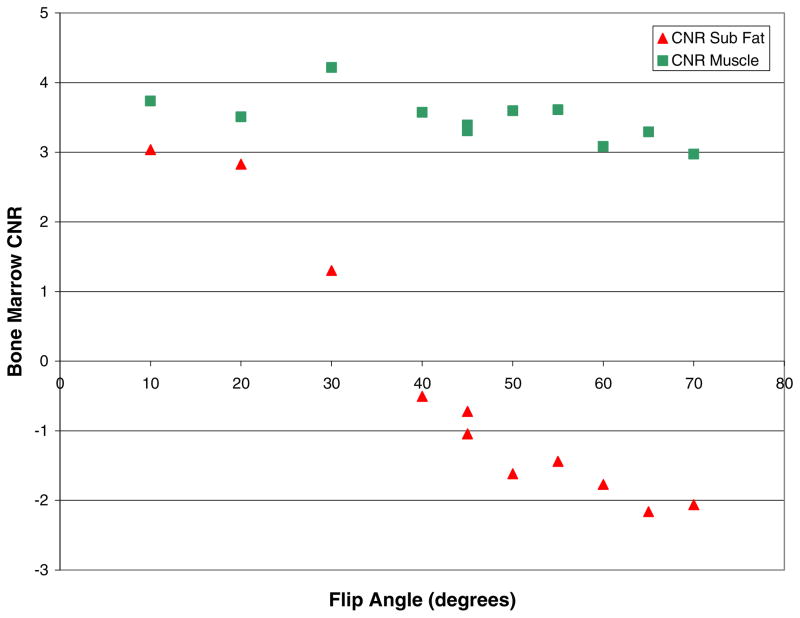
Methods: MR acquisition parameters were first optimized at 3 Tesla. Thereafter, five volunteers underwent axial and coronal exams of their right knee. Each image series was evaluated visually and quantitatively. An anatomically standardized region-of-interest was placed on both the medial and lateral tibial plateaus of all coronal slices containing subchondral bone. Mean and maximum marrow signal was measured, and "bone signal" was calculated.
Results: The MR acquisition had spatial resolution 0.2 × 0.2 × 1.0 mm and acquisition time 10.5 min. The two asymptomatic knees exhibited prominent horizontal trabeculae in the tibial subchondral bone, while the one confirmed OA knee had disorganized subchondral bone and absent horizontal trabeculae. The subchondral bone signal was 8-14% higher in both compartments of the OA knee than the asymptomatic knees.
Conclusion: The weight-bearing femorotibial subchondral trabecular bone can be directly visualized and changes quantified in the coronal-oblique plane. Qualitative and quantitative assessments can be performed using the resultant images and may provide a method to discriminate between the healthy and OA knees. These methods should enable a quantitative evaluation of the role of weight-bearing subchondral bone in the natural history of knee OA to be undertaken.
Conflict of interest statement
Figures


References
-
- Moskowitz RW. Bone remodeling in osteoarthritis: subchondral and osteophytic responses. Osteoarthritis Cartilage. 1999;7:323–4. - PubMed
-
- Lajeunesse D. The role of bone in the treatment of osteoarthritis. Osteoarthritis Cartilage. 2004;12(Suppl A):S34–8. - PubMed
-
- Karsdal MA, Leeming DJ, Dam EB, Henriksen K, Alexandersen P, Pastoureau P, et al. Should subchondral bone turnover be targeted when treating osteoarthritis? Osteoarthritis Cartilage. 2008;16:638–46. - PubMed
-
- Lajeunesse D, Hilal G, Pelletier JP, Martel-Pelletier J. Subchondral bone morphological and biochemical alterations in osteoarthritis. Osteoarthritis Cartilage. 1999;7:321–2. - PubMed
Publication types
MeSH terms
Grants and funding
LinkOut - more resources
Full Text Sources
