

A quantitative structure-activity relationship for translocation of tripeptides via the human proton-coupled peptide transporter, hPEPT1 (SLC15A1)
- PMID: 20449699
- PMCID: PMC2895456
- DOI: 10.1208/s12248-010-9195-z
A quantitative structure-activity relationship for translocation of tripeptides via the human proton-coupled peptide transporter, hPEPT1 (SLC15A1)
Abstract
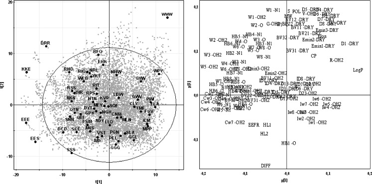
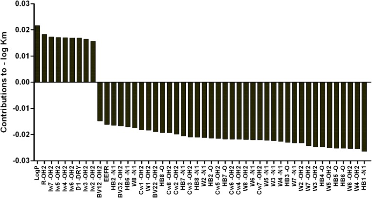
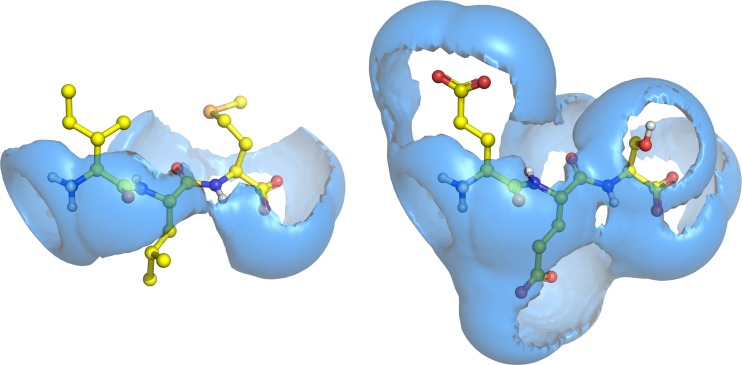
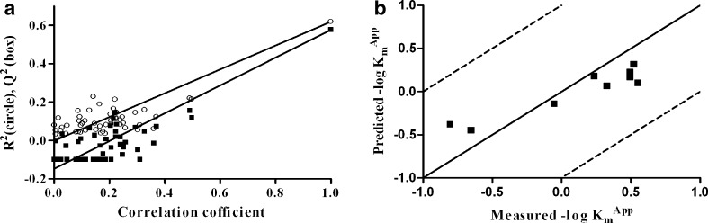
The human intestinal proton-coupled peptide transporter, hPEPT1 (SLC15A1), has been identified as an absorptive transporter for both drug substances and prodrugs. An understanding of the prerequisites for transport has so far been obtained from models based on competition experiments. These models have limited value for predicting substrate translocation via hPEPT1. The aim of the present study was to investigate the requirements for translocation via hPEPT1. A set of 55 tripeptides was selected from a principal component analysis based on VolSurf descriptors using a statistical design. The majority of theses tripeptides have not previously been investigated. Translocation of the tripeptides via hPEPT1 was determined in a MDCK/hPEPT1 cell-based translocation assay measuring substrate-induced changes in fluorescence of a membrane potential-sensitive probe. Affinities for hPEPT1 of relevant tripeptides were determined by competition studies with [14C]Gly-Sar in MDCK/hPEPT1 cells. Forty tripeptides were found to be substrates for hPEPT1, having K(m)(app) values in the range 0.4-28 mM. Eight tripeptides were not able to cause a substrate-induced change in fluorescence in the translocation assay and seven tripeptides interacted with the probe itself. The conformationally restricted tripeptide Met-Pro-Pro was identified as a novel high-affinity inhibitor of hPEPT1. We also discovered the first tripeptide (Asp-Ile-Arg) that was neither a substrate nor an inhibitor of hPEPT1. To rationalise the requirements for transport, a quantitative structure-activity relationship model correlating K(m)(app) values with VolSurf descriptors was constructed. This is, to our knowledge, the first predictive model for the translocation of tripeptides via hPEPT1.
Figures

Similar articles
-
Design, synthesis, and evaluation of tripeptidic promoieties targeting the intestinal peptide transporter hPEPT1.ChemMedChem. 2007 Apr;2(4):479-87. doi: 10.1002/cmdc.200600200. ChemMedChem. 2007. PMID: 17407174
-
Development of a QSAR model for binding of tripeptides and tripeptidomimetics to the human intestinal di-/tripeptide transporter hPEPT1.Pharm Res. 2006 Mar;23(3):483-92. doi: 10.1007/s11095-006-9462-y. Epub 2006 Feb 26. Pharm Res. 2006. PMID: 16489544
-
In vitro evaluation of N-methyl amide tripeptidomimetics as substrates for the human intestinal di-/tri-peptide transporter hPEPT1.Eur J Pharm Sci. 2006 Jul;28(4):325-35. doi: 10.1016/j.ejps.2006.03.007. Epub 2006 Mar 28. Eur J Pharm Sci. 2006. PMID: 16713701
-
Di/tri-peptide transporters as drug delivery targets: regulation of transport under physiological and patho-physiological conditions.Curr Drug Targets. 2003 Jul;4(5):373-88. doi: 10.2174/1389450033491028. Curr Drug Targets. 2003. PMID: 12816347 Review.
-
Di- and tripeptide transport in vertebrates: the contribution of teleost fish models.J Comp Physiol B. 2017 Apr;187(3):395-462. doi: 10.1007/s00360-016-1044-7. Epub 2016 Nov 1. J Comp Physiol B. 2017. PMID: 27803975 Review.
Cited by
-
Current Evidence on the Bioavailability of Food Bioactive Peptides.Molecules. 2020 Sep 29;25(19):4479. doi: 10.3390/molecules25194479. Molecules. 2020. PMID: 33003506 Free PMC article. Review.
-
Salt Bridge Swapping in the EXXERFXYY Motif of Proton-coupled Oligopeptide Transporters.J Biol Chem. 2015 Dec 11;290(50):29931-40. doi: 10.1074/jbc.M115.675603. Epub 2015 Oct 19. J Biol Chem. 2015. PMID: 26483552 Free PMC article.
-
The tandem chain extension aldol reaction used for synthesis of ketomethylene tripeptidomimetics targeting hPEPT1.Bioorg Med Chem Lett. 2011 Aug 1;21(15):4597-601. doi: 10.1016/j.bmcl.2011.05.108. Epub 2011 Jun 6. Bioorg Med Chem Lett. 2011. PMID: 21703856 Free PMC article.
-
Ibuprofen is a non-competitive inhibitor of the peptide transporter hPEPT1 (SLC15A1): possible interactions between hPEPT1 substrates and ibuprofen.Br J Pharmacol. 2010 Dec;161(8):1793-805. doi: 10.1111/j.1476-5381.2010.01000.x. Br J Pharmacol. 2010. PMID: 20726987 Free PMC article.
References
-
- Addison JM, Burston D, Dalrymple JA, Matthews DM, Payne JW, Sleisenger MH, Wilkinson S. A common mechanism for transport of di- and tri-peptides by hamster jejunum in vitro. Clin Sci Mol Med. 1975;49:313–322. - PubMed
Publication types
MeSH terms
Substances
LinkOut - more resources
Full Text Sources
Miscellaneous
