Multicenter Study
. 2008 Nov;36(11 Suppl):S397-404.
doi: 10.1097/ccm.0b013e31818a7e56.

From laboratory science to six emergency medical services systems: New understanding of the physiology of cardiopulmonary resuscitation increases survival rates after cardiac arrest

Affiliations
Multicenter Study

From laboratory science to six emergency medical services systems: New understanding of the physiology of cardiopulmonary resuscitation increases survival rates after cardiac arrest

Tom P Aufderheide et al. Crit Care Med. 2008 Nov.

Abstract

Objective: The purpose of this study is to: 1) describe a newly mechanism of blood flow to the brain during cardiopulmonary resuscitation using the impedance threshold device in a piglet model of cardiac arrest, and 2) describe the survival benefits in humans of applying all of the highly recommended changes in the 2005 guidelines related to increasing circulation during cardiopulmonary resuscitation, including use of the impedance threshold device, from six emergency medical services systems in the United States.

Design: Animal studies prospective trial with each piglet serving as its own control. Historical controls were used for the human studies.

Subjects: Piglets and patients with out-of-hospital cardiac arrest.

Interventions: Piglets (10-12 kg) were treated with an active (n = 9) or sham (n = 9) impedance threshold device after 6 mins of ventricular fibrillation. Humans were treated with cardiopulmonary resuscitation per the American Heart Association 2005 guidelines and the impedance threshold device.

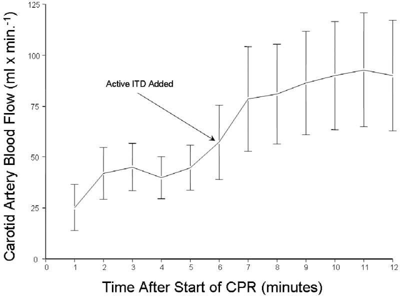
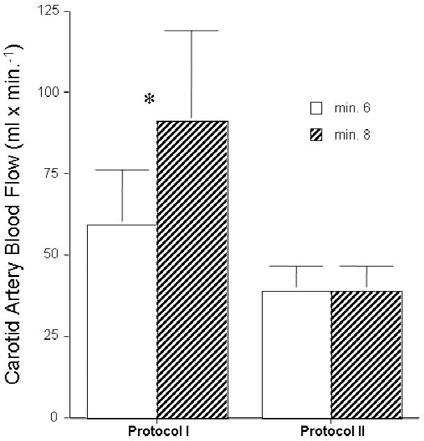
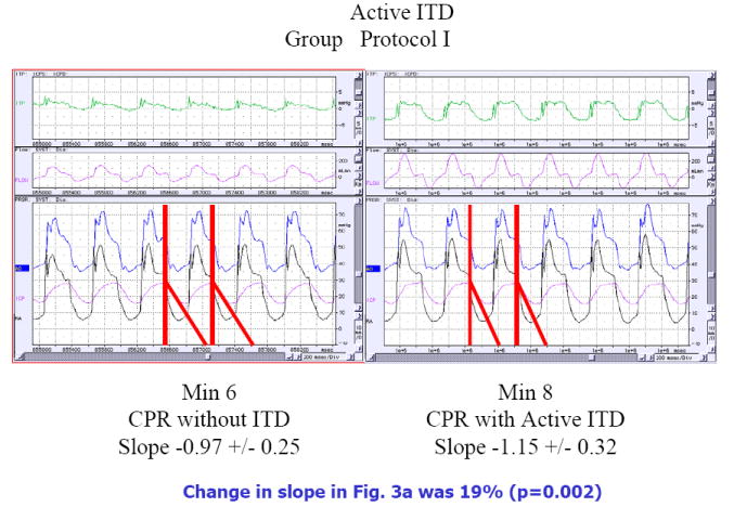
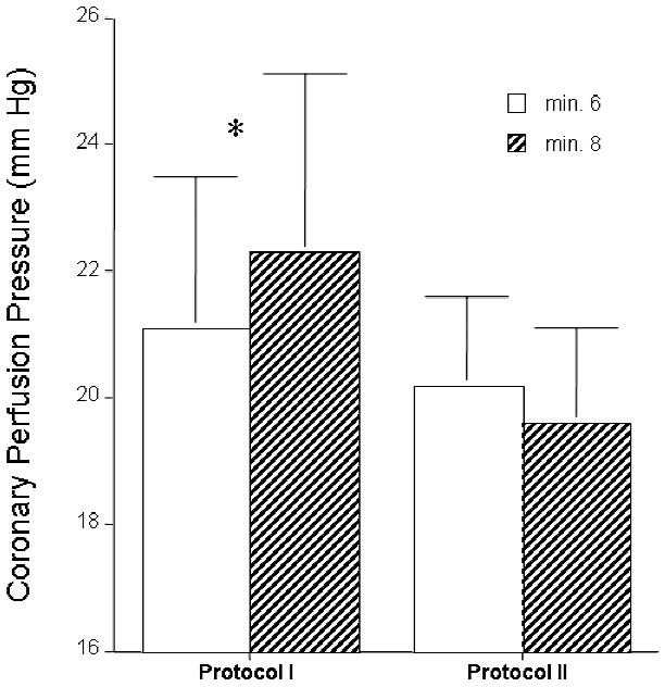
Animals: The primary endpoint in the piglet study was carotid blood flow which increased from 59 mL/min without an impedance threshold device to 91 mL/min (p = 0.017) with impedance threshold device use. Airway pressures during the chest recoil phase decreased from -0.46 mm Hg to -2.59 mm Hg (p = 0.0006) with the active impedance threshold device. Intracranial pressure decreased more rapidly and to a greater degree during the decompression phase of cardiopulmonary resuscitation with the active impedance threshold device. Humans: Conglomerate quality assurance data were analyzed from six emergency medical services systems in the United States serving a population of approximately 3 million people. There were 920 patients treated for cardiac arrest after implementation of the 2005 American Heart Association guidelines, including impedance threshold device use, and 1750 patients in the control group during the year before implementation. Demographics were similar between the two groups. Survival to hospital discharge was 9.3% in the control group versus 13.6% in the intervention group. The odds ratio, 95% confidence interval, and p value were 1.54 (1.19-1.99) and p = 0.0008, respectively. This survival advantage was conferred to patients with a presenting cardiac arrest rhythm of ventricular fibrillation (28.5% vs. 18.0%, p = 0.0008).

Conclusions: Use of the impedance threshold device in piglets increased carotid blood flow and coronary and cerebral perfusion pressures and reduced intracranial pressure during the decompression phase of cardiopulmonary resuscitation at a faster rate than controls, resulting in a longer duration of time when intracranial pressures are at their nadir. Patients in six emergency medical services systems treated with the impedance threshold device together with the renewed emphasis on more compressions, fewer ventilations, and complete chest wall recoil had a nearly 50% increase in survival rates after out-of-hospital cardiac arrest compared with historical controls.


Figures

Figure 1
Figure 1
The means +/- SEM carotid blood flow increased when the ITD was added to the respiratory circuit after 8 minutes of untreated ventricular fibrillation and 6 minutes of CPR without the ITD. The increase was rapid and resulted in a 50% increase in carotid blood flow.
Figure 2
Figure 2
Two groups of piglets were subjected to 8 minutes of untreated ventricular fibrillation followed by 6 minutes of CPR alone and then CPR with either an active or a sham ITD. Carotid blood flow increased after addition of an active ITD in Protocol I but did not change after adding a sham ITD in Protocol II.
Figure 3
Figure 3
Piglets were subjected to 8 minutes of untreated ventricular fibrillation followed by 6 minutes of CPR alone and then CPR with either an active or a sham ITD. The effect of adding an active (Fig 3a) or a sham (Fig 3b) ITD on changes in airway pressures (ITP), carotid blood flow (flow in pink), aortic pressure (Ao in blue), right atrial pressure (RA in black), and intracranial pressure (ICP in pink) are shown in these representative figures. The data is shown after 6 minute of CPR alone and then at minute 8, 2 minutes after addition of the active ITD (Figure 3a) or a sham ITD (Fig 3b). The change in the slope of the ICP curves are highlighted by the red line and the actual values are shown below each figure. When the active ITD is added to the respiratory circuit there is a more rapid decrease in the slope of the ICP curve during the decompression phase of CPR resulting in less resistance to forward blood flow for a longer period of time (Fig 3a). This effect was not observed with the addition of the sham ITD in Fig 3b). These figures also highlight the decreases observed during the chest wall recoil phase of the airway pressure curves with the addition of the active ITD (labeled ITP for intrathoracic pressure in green) in the top panels. This effect is absent with the sham ITD.
Figure 3
Figure 3
Piglets were subjected to 8 minutes of untreated ventricular fibrillation followed by 6 minutes of CPR alone and then CPR with either an active or a sham ITD. The effect of adding an active (Fig 3a) or a sham (Fig 3b) ITD on changes in airway pressures (ITP), carotid blood flow (flow in pink), aortic pressure (Ao in blue), right atrial pressure (RA in black), and intracranial pressure (ICP in pink) are shown in these representative figures. The data is shown after 6 minute of CPR alone and then at minute 8, 2 minutes after addition of the active ITD (Figure 3a) or a sham ITD (Fig 3b). The change in the slope of the ICP curves are highlighted by the red line and the actual values are shown below each figure. When the active ITD is added to the respiratory circuit there is a more rapid decrease in the slope of the ICP curve during the decompression phase of CPR resulting in less resistance to forward blood flow for a longer period of time (Fig 3a). This effect was not observed with the addition of the sham ITD in Fig 3b). These figures also highlight the decreases observed during the chest wall recoil phase of the airway pressure curves with the addition of the active ITD (labeled ITP for intrathoracic pressure in green) in the top panels. This effect is absent with the sham ITD.
Figure 4
Figure 4
Piglets were subjected to 8 minutes of untreated ventricular fibrillation followed by 6 minutes of CPR alone and then CPR with either an active or a sham ITD and the coronary (Fig 4a) and cerebral (Fig 4b) perfusion pressures were compared. Adding an active ITD (Fig 4a) resulted in a statistically significant increase in coronary perfusion pressures (calculated by subtracting the right atrial pressure from the aortic pressure during the decompression phase of CPR). By contrast, coronary perfusion pressures trended lower after adding a sham ITD (Fig 4a). Similarly, adding an active ITD resulted in a statistically significant increase in cerebral perfusion pressures (Fig 4b) (difference between mean arterial pressure and intracranial pressure) whereas addition of the sham ITD had no benefit.
Figure 4
Figure 4
Piglets were subjected to 8 minutes of untreated ventricular fibrillation followed by 6 minutes of CPR alone and then CPR with either an active or a sham ITD and the coronary (Fig 4a) and cerebral (Fig 4b) perfusion pressures were compared. Adding an active ITD (Fig 4a) resulted in a statistically significant increase in coronary perfusion pressures (calculated by subtracting the right atrial pressure from the aortic pressure during the decompression phase of CPR). By contrast, coronary perfusion pressures trended lower after adding a sham ITD (Fig 4a). Similarly, adding an active ITD resulted in a statistically significant increase in cerebral perfusion pressures (Fig 4b) (difference between mean arterial pressure and intracranial pressure) whereas addition of the sham ITD had no benefit.

Similar articles

Cited by

References

    1. Kouwenhoven WB, Jude JR, Knickerbocker GG. Closed chest cardiac massage. JAMA. 1960;173:1064–1067. - PubMed
    1. Duggal C, Weil MH, Gazmuri RJ, Tang W, Sun S, O’Connell F, Ali M. Regional blood flow during closed-chest cardiac resuscitation in rats. J Appl Physiol. 1993 Jan;74(1):147–52. - PubMed
    1. Andreka P, Frenneaux MP. Haemodynamics of cardiac arrest and resuscitation. Curr Opin Crit Care. 2006 Jun;12(3):198–203. - PubMed
    1. Niemann JT. Cardiopulmonary resuscitation. N Engl J Med. 1992;327:1075–1090. - PubMed
    1. Eisenberg MS, Horwood BT, Cummins RO, et al. Cardiac arrest and resuscitation: a tale of 29 cities. Ann Emerg Med. 1990;19:179–186. - PubMed

Publication types

MeSH terms