Review
. 2008 Nov;36(11 Suppl):S440-6.
doi: 10.1097/ccm.0b013e31818a89f4.

Targeting mitochondria for resuscitation from cardiac arrest

Affiliations
Review

Targeting mitochondria for resuscitation from cardiac arrest

Iyad M Ayoub et al. Crit Care Med. 2008 Nov.

Abstract

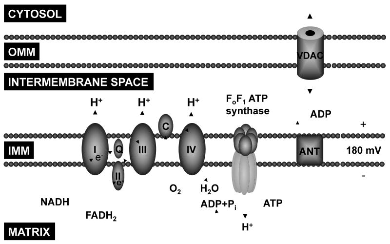
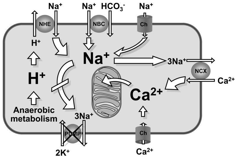
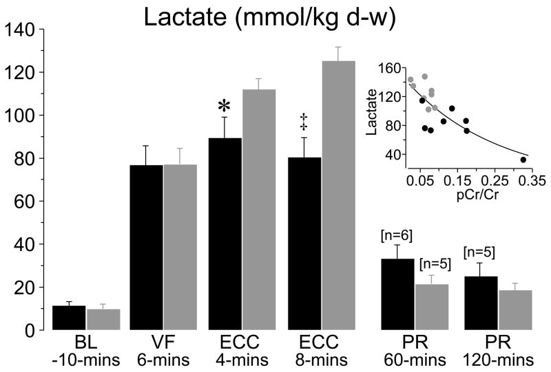
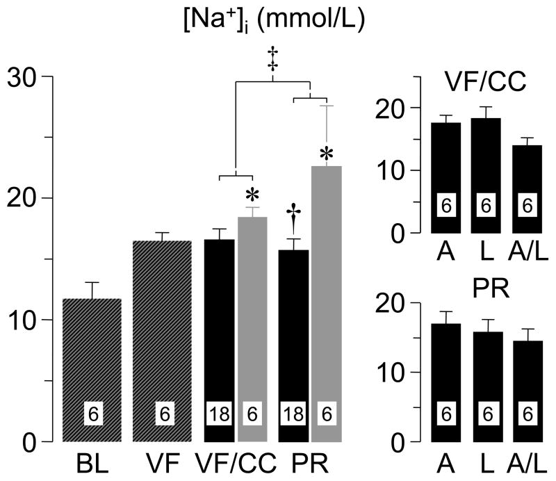
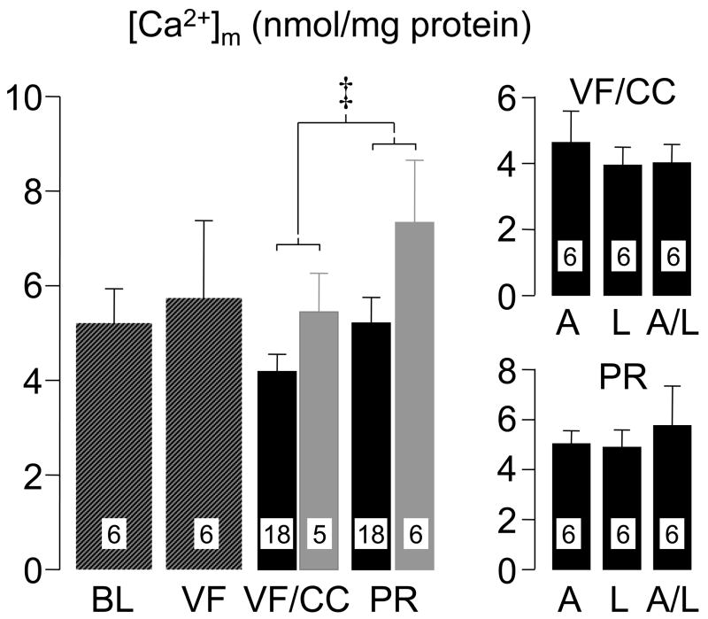
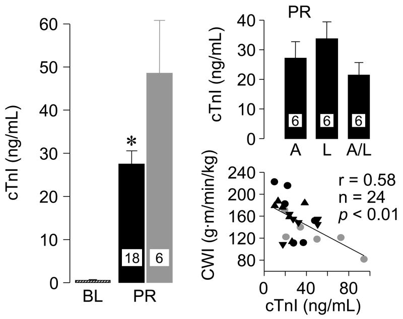
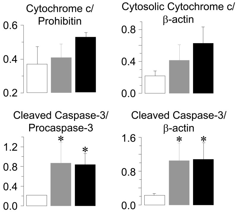
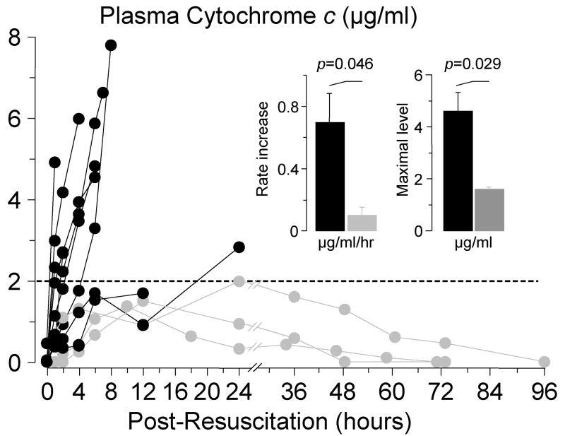
Reversal of cardiac arrest requires reestablishment of aerobic metabolism by reperfusion with oxygenated blood of tissues that have been ischemic for variable periods of time. However, reperfusion concomitantly activates a myriad of pathogenic mechanisms causing what is known as reperfusion injury. At the center of reperfusion injury are mitochondria, playing a critical role as effectors and targets of injury. Studies in animal models of ventricular fibrillation have shown that limiting myocardial cytosolic Na+ overload attenuates mitochondrial Ca2+ overload and maintains oxidative phosphorylation, which is the main bioenergetic function of mitochondria. This effect is associated with functional myocardial benefits such as preservation of myocardial compliance during chest compression and attenuation of myocardial dysfunction after return of spontaneous circulation. Additional studies in similar animal models of ventricular fibrillation have shown that mitochondrial injury leads to activation of the mitochondrial apoptotic pathway, characterized by the release of cytochrome c to the cytosol, reduction of caspase-9 levels, and activation of caspase-3 coincident with marked reduction in left ventricular function. Cytochrome c also "leaks" into the bloodstream attaining levels that are inversely proportional to survival. These data indicate that mitochondria play a key role during cardiac resuscitation by modulating energy metabolism and signaling apoptotic cascades and that targeting mitochondria could represent a promising strategy for cardiac resuscitation.


Figures

Figure 1
Figure 1
Schematic rendition of key mitochondrial components involved in ATP synthesis via oxidative phosphorylation. OMM, outer mitochondrial membrane; IMM, inner mitochondrial membrane; I, II, III, and IV, respiratory chain complexes; Q, coenzyme Q; C, cytochrome c; ANT, adenine nucleotide translocator; VDAC, voltage-dependent anion channel.
Figure 2
Figure 2
Schematic rendition of a cardiomyocyte during ischemia and reperfusion depicting Na+-induced cytosolic and mitochondrial Ca2+ overload. NHE, sodium-hydrogen exchanger iso-form-1; NBC, Na+-HCO3 cotransporter; NCX, Na+-Ca2+ exchanger; Ch, channel.
Figure 3
Figure 3
Diagram depicting an open chest pig model of ventricular fibrillation and extracorporeal circulation. ECC, system for extracorporeal circulation; PPV, positive pressure ventilation; RA, right atrial pressure; CO, cardiac output; MAP, mean aortic pressure; ECG, electrocardiogram; LAD, left anterior descending coronary artery; LV, left ventricle; HEM (HPLC), high energy metabolites analyzed by high performance liquid chromatography. (Adapted from Ayoub I et al. Crit Care Med 2007;35:2329–36) (49).
Figure 4
Figure 4
Creatine phosphate/creatine (PCr/Cr) ratios, adenosine triphosphate/adenosine diphosphate (ATP/ADP) ratios, and adenosine levels measured in left ventricular tissue in pigs randomized to receive either 3 mg/kg of zoniporide (black bars) or 0.9% NaCl (gray bars) into the right atrium after 8 minutes of untreated ventricular fibrillation, immediately before starting extracorporeal circulation (ECC). d-w, dry-weight. Measurements were obtained at baseline (BL), at minute 4 of ECC (ECC 4), at minute 8 of ECC (ECC 8), and at 60 minutes post-resuscitation (PR). Each group included 8 pigs each at baseline and during ECC and 6 pigs in the zoniporide group and 5 in the NaCl group at post-resuscitation. Values are mean ± SEM. Differences were tested by Student’s t-test. (Adapted from Ayoub I et al. Crit Care Med 2007;35:2329–36) (49).
Figure 5
Figure 5
Myocardial lactate levels in left ventricular tissue in pigs randomized to receive either zoniporide (black symbols, n = 8) or NaCl (gray symbols, n = 8) before extracorporeal circulation. Numbers in brackets indicate when sample size decreased from the initial eight or from the preceding sample size. Insert demonstrates the relationship between myocardial lactate and the creatine phosphate to creatine ratio (pCr/Cr) at ECC 8 minutes. The regression line represents an exponential decay function (R2 = 0.63, p < 0.001). BL, baseline; VF, ventricular fibrillation; ECC, extracorporeal circulation; PR, postresuscitation; d-w, dry weight. Values are mean ± SEM; *p < 0.05, ‡p < 0.001 vs. NaCl by Student’s t-test. (Adapted from Ayoub I et al. Crit Care Med 2007;35:2329–36) (49).
Figure 6
Figure 6
Intracellular Na+ ([Na+]i) in left ventricular tissue of rats at baseline (BL), at 15 minutes of untreated ventricular fibrillation (VF), at 15 minutes of VF accompanied by 5 minutes of chest compression (CC), and at 60 minutes post-resuscitation (PR). Hatched bars represent measurements without pharmacological treatment. Black bars represent rats treated with Na+ limiting interventions. Gray bars represent rats treated with vehicle control. The individual Na+-limiting interventions are shown on the right panels; A, selective sodium-hydrogen exchanger isoform-1 inhibitor AVE4454; L, lidocaine; and A/L, combination of AVE4454 and lidocaine. Numbers within bars denote number of hearts processed for the measurement. Values are mean ± SEM. *p < 0.05 vs BL by Kruskal-Wallis one-way ANOVA on ranks using Dunn’s Method for multiple comparisons; p < 0.05 vs control by Student’s t-test in PR groups; ‡two-way ANOVA using time factor (VF/CC vs PR) and treatment factor (control vs Na+-limiting interventions) was significant for treatment factor (p = 0.013). (Adapted from Wang S et al. J Appl Physiol 2007;103:55–65) (48).
Figure 7
Figure 7
Mitochondrial Ca2+ ([Ca2+]m) in left ventricular tissue of rats. For interpretation of bars and abbreviations refer to legend for Figure 6. Two-way ANOVA using time factor (VF/CC vs PR) and treatment factor (control vs Na+-limiting interventions) was significant for both, time factor (p = 0.045) and treatment factor (p = 0.021). (Adapted from Wang S et al. J Appl Physiol 2007;103:55–65) (48).
Figure 8
Figure 8
Cardiospecific troponin I (cTnI) in plasma at baseline (BL) and at 60 minutes post-resuscitation (PR) in rats subjected to ventricular fibrillation and resuscitation. Black symbols represent rats treated with a Na+-limiting intervention (AVE4454 circles, lidocaine inverted triangle, and AVE4454 and lidocaine combined upright triangles). Gray symbols represent control rats. Values are mean ± SEM. *P< 0.05 vs. control by Student’s t-test. The scatterplot depicts the correlation between cTnI and cardiac work index (CWI) at 60 min post-resuscitation. (Adapted from Wang S et al. J Appl Physiol 2007;103:55–65) (48).
Figure 9
Figure 9
Densitometry of left ventricular immunoblots demonstrating numerical increases in mitochondrial cytochrome c relative to prohibitin and cytosolic cytochrome c relative to β-actin and statistically significant increases in 17 kDa cleaved caspase-3 fragments in the cytosolic fraction relative to pro-caspase-3 and β-actin at 240 minutes post-resuscitation. Rats were randomized to untreated VF lasting 4 minutes (gray bars, n = 4), 8 minutes (black bars, n = 4), or to sham intervention (open bars, n = 4). Values are mean ± SEM. *p < 0.05 vs sham by one-way ANOVA and Dunn’s test for multiple comparisons. (Adapted from Radhakrishnan J et al. Am J Physiol Heart Circ Physiol 2007; 292:767–75) (53).
Figure 10
Figure 10
Serial measurements of plasma cytochrome c by reverse-phase high performance liquid chromatography in rats successfully resuscitated after 8 minutes of untreated ventricular fibrillation. Measurements were made until cytochrome c levels had returned to baseline or the rat had died. Gray symbols represent survivors (n = 3); black symbols represent non-survivors (n = 9). (Adapted from Radhakrishnan J et al. Am J Physiol Heart Circ Physiol 2007; 292:767–75) (53).

Similar articles

Cited by

References

    1. Rosamond W, Flegal K, Furie K, Go A, Greenlund K, Haase N, Hailpern SM, Ho M, Howard V, Kissela B, Kittner S, Lloyd-Jones D, McDermott M, Meigs J, Moy C, Nichol G, O’Donnell C, Roger V, Sorlie P, Steinberger J, Thom T, Wilson M, Hong Y. Heart disease and stroke statistics--2008 update: a report from the American Heart Association Statistics Committee and Stroke Statistics Subcommittee. Circulation. 2008;117:e25–146. - PubMed
    1. Sans S, Kesteloot H, Kromhout D. The burden of cardiovascular diseases mortality in Europe. Task Force of the European Society of Cardiology on Cardiovascular Mortality and Morbidity Statistics in Europe. Eur Heart J. 1997;18:1231–1248. - PubMed
    1. Brown CG, Martin DR, Pepe PE, Stueven H, Cummins RO, Gonzalez E, Jastremski M the Multicenter High-Dose Epinephrine Study Group. A comparison of standard-dose and high-dose epinephrine in cardiac arrest outside the hospital. N Engl J Med. 1992;327:1051–1055. - PubMed
    1. Kellermann AL, Hackman BB, Somes G. Predicting the outcome of unsuccessful prehospital advanced cardiac life support. JAMA. 1993;270:1433–1436. - PubMed
    1. Lombardi G, Gallagher J, Gennis P. Outcome of out-of-hospital cardiac arrest in New York City. The pre-hospital arrest survival evaluation (PHASE) study. JAMA. 1994;271:678–683. - PubMed

MeSH terms