Correction of axial and rotational alignment after medial and lateral releases during balanced gap TKA. A clinical study of 54 patients
- PMID: 20450421
- PMCID: PMC2876838
- DOI: 10.3109/17453674.2010.483992
Correction of axial and rotational alignment after medial and lateral releases during balanced gap TKA. A clinical study of 54 patients
Abstract
Background and purpose: Restoration of mechanical alignment after total knee arthroplasty can be achieved with ligament releases. Several previously described sequences and results achieved with cadaver knees, with measured resection implantation techniques, may not be applied to the balanced gap technique. We investigated the peroperative effect of stepwise soft tissue releases following the "tightest structure first" on leg axis in extension and femur rotation in flexion.
Methods: During PCL-retaining total knee arthroplasty (TKA), using a balanced gap technique in 54 patients we determined the effect of each ligament release using a navigation system while the knee was distracted with a tensor in extension and flexion. The effect on alignment in extension and on femoral rotation in flexion was measured for each release separately.
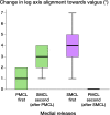
Results: In more than half of the patients, one or more ligament releases were necessary. Release of the posteromedial condyle led to a minor effect on leg axis in extension and femoral rotation in flexion, release of the superficial medial collateral ligament to a few degrees, mainly in extension. Release of the iliotibial tract led to a small correction of leg alignment in extension. There was no statistically significant difference in the alignment-correcting effect of a release dependent upon the sequence in which the structure was released.
Interpretation: In PCL-retaining TKA, a stepwise "tightest structure first" protocol for ligament releases in extension with the balanced gap technique results in effective, gradual, alignment correction in extension, and limited femoral rotating effects in flexion.
Figures


References
-
- Berger A, Rubash E, Seel J, Thompson H, Crossett S. Determining the rotational alignment of the femoral component in total knee arthroplasty using the epicondylar axis. Clin Orthop. 1993;((286)):40–7. - PubMed
-
- Clarke HD, Fuchs R, Scuderi GR, Scott WN, Insall JN. Clinical results in valgus total knee arthroplasty with the ”pie crust” technique of lateral soft tissue releases. J Arthroplasty. 2005;20:1010–4. - PubMed
-
- Fehring TK. Rotational malalignment of the femoral component in total knee arthroplasty. Clin Orthop. 2000;((380)):72–9. - PubMed
-
- Fehring TK, Odum S, Griffin WL, Mason JB, Nadaud M. Early failures in total knee arthroplasty. Clin Orthop Relat Res. 2001;((392)):315–8. - PubMed
Publication types
MeSH terms
LinkOut - more resources
Full Text Sources
Medical
Miscellaneous
