

Appearance of the weight-bearing lateral radiograph in retrocalcaneal bursitis
- PMID: 20450438
- PMCID: PMC2876845
- DOI: 10.3109/17453674.2010.487245
Appearance of the weight-bearing lateral radiograph in retrocalcaneal bursitis
Abstract
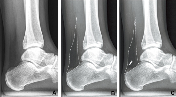
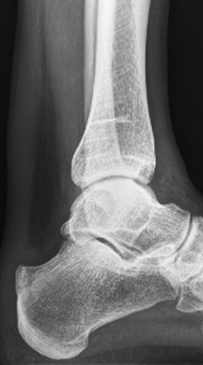
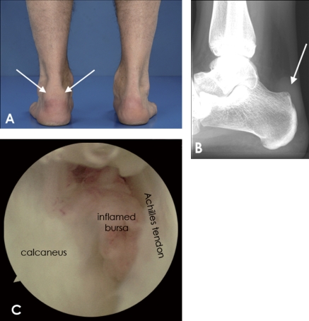
Background and purpose: A retrocalcaneal bursitis is caused by repetitive impingement of the bursa between the Achilles tendon and the posterosuperior calcaneus. The bursa is situated in the posteroinferior corner of Kager's triangle (retrocalcaneal recess), which is a radiolucency with sharp borders on the lateral radiograph of the ankle. If there is inflammation, the fluid-filled bursa is less radiolucent, making it difficult to delineate the retrocalcaneal recess. We assessed whether the radiographic appearance of the retrocalcaneal recess on plain digital (filmless) radiographs could be used in the diagnosis of a retrocalcaneal bursitis.
Methods: Whether or not there was obliteration of the retrocalcaneal recess (yes/no) on 74 digital weight-bearing lateral radiographs of the ankle was independently assessed by 2 observers. The radiographs were from 24 patients (25 heels) with retrocalcaneal bursitis (confirmed on endoscopic calcaneoplasty); the control group consisted of 50 patients (59 heels).
Results: The sensitivity of the test was 83% for observer 1 and 79% for observer 2. Specificity was 100% and 98%, respectively. The kappa value of the interobserver reliability test was 0.86. For observer 1, intraobserver reliability was 0.96 and for observer 2 it was 0.92.
Interpretation: On digital weight-bearing lateral radiographs of a retrocalcaneal bursitis, the retrocalcaneal recess has a typical appearance.
Figures
Similar articles
-
The appearance of the pre-Achilles fat pad after endoscopic calcaneoplasty.Knee Surg Sports Traumatol Arthrosc. 2015 Aug;23(8):2400-2405. doi: 10.1007/s00167-014-2908-6. Epub 2014 Mar 1. Knee Surg Sports Traumatol Arthrosc. 2015. PMID: 24584645
-
High patient satisfaction and good long-term functional outcome after endoscopic calcaneoplasty in patients with retrocalcaneal bursitis.Knee Surg Sports Traumatol Arthrosc. 2021 May;29(5):1494-1501. doi: 10.1007/s00167-020-06167-2. Epub 2020 Jul 25. Knee Surg Sports Traumatol Arthrosc. 2021. PMID: 32712686 Free PMC article.
-
The retrocalcaneal bursa: anatomy and bursography.Foot Ankle. 1992 May;13(4):203-7. doi: 10.1177/107110079201300407. Foot Ankle. 1992. PMID: 1634153
-
Histo-Anatomy and Sonographic Examination for the Retrocalcaneal Bursal Complex: EURO-MUSCULUS/USPRM Approach.J Ultrasound Med. 2024 Nov;43(11):2027-2038. doi: 10.1002/jum.16544. Epub 2024 Aug 13. J Ultrasound Med. 2024. PMID: 39136225 Review.
-
Minimally Invasive and Endoscopic Treatment of Haglund Syndrome.Foot Ankle Clin. 2019 Sep;24(3):515-531. doi: 10.1016/j.fcl.2019.04.006. Epub 2019 May 18. Foot Ankle Clin. 2019. PMID: 31371001 Review.
Cited by
-
Anatomical and clinical relevance of Kager's triangle: diagnostic approaches and therapeutic implications for related pathologies.J Orthop Surg Res. 2025 Jul 19;20(1):682. doi: 10.1186/s13018-025-06081-8. J Orthop Surg Res. 2025. PMID: 40684192 Free PMC article. Review.
-
Haglund's Syndrome: endoscopic or open treatment?Acta Biomed. 2020 May 30;91(4-S):167-171. doi: 10.23750/abm.v91i4-S.9576. Acta Biomed. 2020. PMID: 32555092 Free PMC article.
-
Characteristics of Pressure on the Apophysis in the Course of Paediatric Heel Pain-Preliminary Report.Int J Environ Res Public Health. 2023 Apr 5;20(7):5403. doi: 10.3390/ijerph20075403. Int J Environ Res Public Health. 2023. PMID: 37048018 Free PMC article.
-
Endoscopic surgery of the Achilles tendon.Curr Rev Musculoskelet Med. 2012 Jun;5(2):156-63. doi: 10.1007/s12178-012-9115-1. Curr Rev Musculoskelet Med. 2012. PMID: 22354353 Free PMC article.
-
The appearance of the pre-Achilles fat pad after endoscopic calcaneoplasty.Knee Surg Sports Traumatol Arthrosc. 2015 Aug;23(8):2400-2405. doi: 10.1007/s00167-014-2908-6. Epub 2014 Mar 1. Knee Surg Sports Traumatol Arthrosc. 2015. PMID: 24584645
References
-
- DeVries JG, Summerhays B, Guehlstorf DW. Surgical correction of Haglund's triad using complete detachment and reattachment of the Achilles tendon. J Foot Ankle Surg. 2009;48:447–51. - PubMed
-
- Fischer E. Soft tissue diagnosis on the extremities using soft tissue radiography. Part I. Indications and some technical aspects of low KeV radiography (author's transl) Radiologe. 1974a;14:454–6. - PubMed
-
- Fischer E. Soft tissue diagnosis on the extremities using soft tissue radiography. Part II. Diseases of the Achilles tendon and the surrounding tissues (author's transl) Radiologe. 1974b;14:457–67. - PubMed
-
- Goodman LR, Shanser JD. The pre-Achilles fat pad: An aid to early diagnosis of local or systemic disease. Skeletal Radiol. 1997;2:81–6.
-
- Heneghan MA, Pavlov H. The Haglund painful heel syndrome. Experimental investigation of cause and therapeutic implications. Clin Orthop. 1984:228–34. - PubMed
MeSH terms
LinkOut - more resources
Full Text Sources
Medical