. 2010 Jul 15;429(2):391-401.
doi: 10.1042/BJ20100336.

The identification of the SNARE complex required for the fusion of VLDL-transport vesicle with hepatic cis-Golgi

Affiliations

The identification of the SNARE complex required for the fusion of VLDL-transport vesicle with hepatic cis-Golgi

Shaila Siddiqi et al. Biochem J. .

Abstract

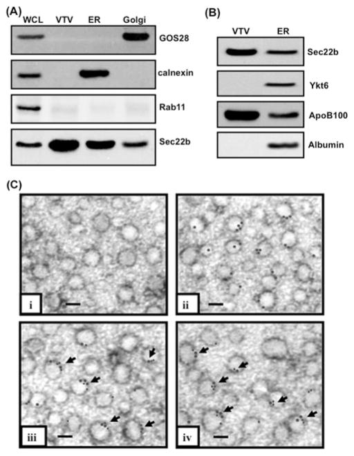
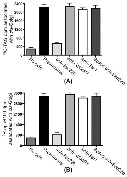
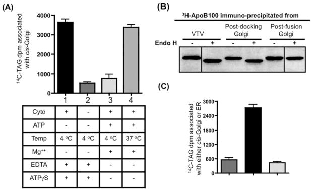
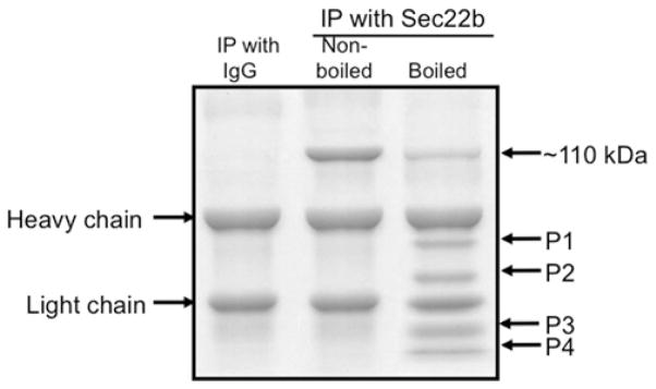
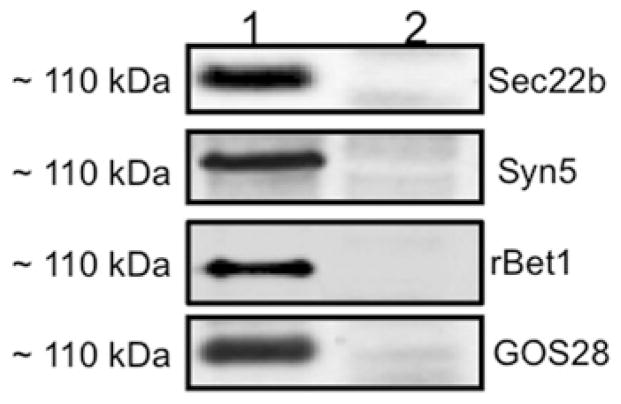
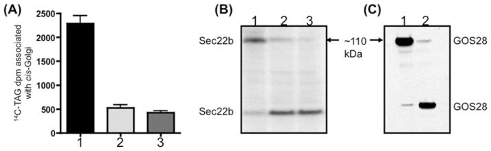
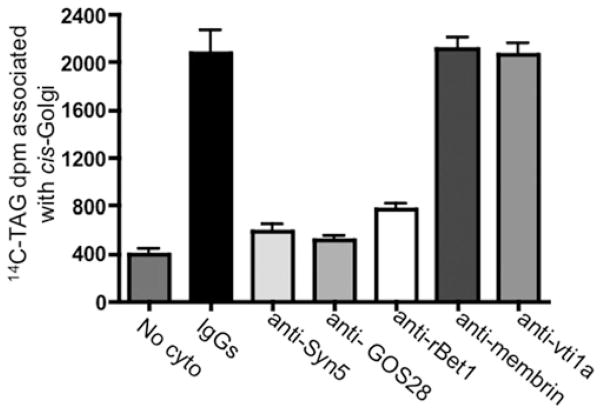
VLDLs (very-low-density lipoproteins) are synthesized in the liver and play an important role in the pathogenesis of atherosclerosis. Following their biogenesis in hepatic ER (endoplasmic reticulum), nascent VLDLs are exported to the Golgi which is a physiologically regulatable event. We have previously shown that a unique ER-derived vesicle, the VTV (VLDL-transport vesicle), mediates the targeted delivery of VLDL to the Golgi lumen. Because VTVs are different from other ER-derived transport vesicles in their morphology and biochemical composition, we speculated that a distinct set of SNARE (soluble N-ethylmaleimide-sensitive factor-attachment protein receptor) proteins would form a SNARE complex which would eventually facilitate the docking/fusion of VTVs with Golgi. Our results show that Sec22b is concentrated in VTVs as compared with the ER. Electron microscopic results show that Sec22b co-localizes with p58 and Sar1 on the VTV surface. Pre-treatment of VTV with antibodies against Sec22b inhibited VTV-Golgi fusion, indicating its role as a v-SNARE (vesicle SNARE). To isolate the SNARE complex, we developed an in vitro docking assay in which VTVs were allowed to dock with the Golgi, but fusion was prevented to stabilize the SNARE complex. After the docking reaction, VTV-Golgi complexes were collected, solubilized in 2% Triton X-100 and the SNARE complex was co-immunoprecipitated using anti-Sec22b or GOS28 antibodies. A approximately 110 kDa complex was identified in non-boiled samples that was dissociated upon boiling. The components of the complex were identified as Sec22b, syntaxin 5, rBet1 and GOS28. Antibodies against each SNARE component significantly inhibited VTV-Golgi fusion. We conclude that the SNARE complex required for VTV-Golgi fusion is composed of Sec22b, syntaxin 5, rBet1 and GOS28.


Figures

Figure 1
Figure 1. Sec22b concentrates on VTVs and co-localizes with p58 and Sar1 on their surface
(A) Samples containing hepatic whole-cell lysate (WCL), VTVs, ER and Golgi (each containing30 μg of protein) were separated by SDS/PAGE (12 % gel), transblotted on to a nitrocellulose membrane and probed with specific antibodies against GOS28, calnexin, and Rab11. For details, see the Experimental section. Protein detection was by ECL. (B) Samples of ER and VTVs (30 μg of protein each) were separated by SDS/PAGE (5–15 % gel), transblotted on to a nitrocellulose membrane and probed with specific antibodies against Sec22b, Ykt6, ApoB100 and albumin. Protein detection was by ECL. (C) Immunoelectron microscopy of VTVs utilizing the negative-staining technique. VTVs were adsorbed on formvar-carbon-coated nickel grids and were treated with: (i) anti-rabbit pre-immune IgG; (ii) rabbit polyclonal anti-Sec22b antibodies detected with anti-(rabbit IgG) labelled with 15 nm gold particles; (iii) mouse anti-Sec22b antibodies detected with anti-(mouse IgG) labelled with 15 nm gold particles and anti-Sar1 antibodies detected with anti-(rabbit IgG) labelled with 10 nm gold particles (arrows show co-localization of Sec22b with Sar1); and (iv) anti-p58 antibodies detected with anti-(rabbit IgG) labelled with 10 nm gold particles and mouse anti-Sec22b antibodies detected with anti-(mouse IgG) labelled with 15 nm gold particles (arrows show co-localization of Sec22b with p58). Scale bars, 100 nm.
Figure 2
Figure 2. VTVs utilize Sec22b as a functional v-SNARE to fuse with hepatic cis-Golgi
VTVs (150 μg of protein) containing [14C]TAG and [3H]apoB100 were incubated at 4 °C with 10 μl of pre-immune IgG, 10 μl of anti-Sec22b antibodies, 10 μl of anti-VAMP7 antibodies, 10 μl of anti-Sar1 antibodies or 10 μl of boiled anti-Sec22b antibodies. Antibodies in each case were removed by washing, and the treated VTVs were incubated with non-radiolabelled cis-Golgi (300 μg of protein) at 37 °C in the presence or absence (no cyto) of cytosol (0.5 mg of protein). (A) After incubation, Golgi membranes were separated and the Golgi-associated [14C]TAG was extracted (A) or the Golgi-associated [3H]apoB100 was immunoprecipitated (B). Total [14C]TAG (A) or [3H]apoB100 (B) levels (in d.p.m.) were determined. For details, see the Experimental section. Results are means + S.E.M. (n = 4).
Figure 3
Figure 3. VTVs dock at hepatic cis-Golgi and require cytosol for the docking and fusion process
(A) VTVs (150 μg of protein) containing [14C]TAG and [3H]apoB100 were incubated with non-radiolabelled cis-Golgi (300 μg of protein) with or without cytosol at either 4 or 37 °C in the presence or absence of Mg2+ and ATP as indicated. After incubation for 30 min, the cis-Golgi fraction was isolated on a sucrose step gradient and collected by aspiration of the 0.86/1.15 M interface, and the amount of [14C]TAG was measured. The Golgi was separated from VTVs and the Golgi-associated [14C]TAG was extracted and levels were measured. Results are means + S.E.M. (n = 4). ATPγS, adenosine 5′-[γ-thio]triphosphate. (B) Exactly the same experiment was carried out as in (A) and the [3H]apoB100 was immunoprecipitated either from the VTVs, post-docking Golgi (A, bar 1) or the post-fusion Golgi (A, bar 4). Immunoprecipitated [3H]apoB100 was incubated with endo H (500 units) for 20 h at 37 °C, separated by SDS/PAGE (5–15 % gel), and autoradiographed. (C) VTVs (150 μg of protein) containing [14C]TAG were incubated with either non-radiolabelled cis-Golgi (300 μg of protein) (left-hand and middle bars) or non-radiolabelled hepatic ER (right-hand bar) at 4 °C in the presence (middle and right-hand bars) or absence (left-hand bar) of cytosol. Mg2+-ATP were excluded in each case. After incubation, the cis-Golgi fraction was isolated and the [14C]TAG levels were determined. Results are means + S.E.M. (n = 4).
Figure 4
Figure 4. Sec22b pulls down a ~110 kDa protein complex from post-docking VTV–Golgi complexes
VTVs (150 μg) were incubated at 4 °C with hepatic cis-Golgi (300 μg), cytosol (500 μg of protein) and without Mg2+-ATP. The VTV–Golgi complexes were isolated on a sucrose step gradient. The VTV–Golgi complexes were solubilized in 2 % (v/v) Triton X-100 and incubated with either pre-immune IgG or anti-Sec22b antibodies bound to agarose beads at 4 °C overnight. The beads were washed and either boiled or not, as indicated, in Laemmli buffer. The proteins were separated by SDS/PAGE (5–15 % gel) and the gel was stained. For further details, see the Experimental section. IP, immunoprecipitation.
Figure 5
Figure 5. Proteins co-immunoprecipitated with Sec22b in a ~110 kDa complex after VTV–Golgi docking
(A) VTVs (150 μg) were incubated at 4 °C with hepatic cis-Golgi (300 μg), cytosol (500 μg of protein), and without Mg2+-ATP. VTV–Golgi complexes were isolated on a sucrose step gradient. VTV–Golgi complexes were solubilized in 2 % (v/v) Triton X-100 and incubated with anti-Sec22b antibodies bound to agarose beads at 4 °C overnight. The beads were washed and either not boiled (lane 1) or boiled (lane 2) in Laemmli buffer. The proteins were separated by SDS/PAGE (5–15 %) and probed with antibodies against the indicated proteins. A single membrane was used, which was sequentially probed with the indicated antibodies after washing. Only protein bands that migrated at ~ 110 kDa are shown. Detection was carried out by ECL. The results are representative of four experiments. (B) Samples of hepatic whole-cell lysate (WCL), VTVs, ER and Golgi (each containing 30 μg of protein) were separated by SDS/PAGE (12 % gel), transblotted on to a nitrocellulose membrane and probed with specific antibodies against the indicated proteins. Protein detection was by ECL.
Figure 6
Figure 6. Proteins co-immunoprecipitated with GOS28 in a ~110 kDa complex after VTV–Golgi docking
A similar VTV–Golgi docking assay was performed as in Figure 5(A), and the VTV–Golgi complexes were isolated. The VTV–Golgi complexes were solubilized in 2 % (v/v) Triton X-100 and incubated with either pre-immune IgG or anti-GOS28 antibodies bound to agarose beads at 4 °C overnight. The beads were washed and either not boiled (lane 1) or boiled (lane 2) in Laemmli buffer. The proteins were separated by SDS/PAGE (5–15 %) and probed with antibodies against the indicated proteins. A single membrane was used, which was sequentially probed with the indicated antibodies after washing. Only protein bands migrated at ~ 110 kDa are shown. Detection was carried out by ECL. The results are representative of four independent trials.
Figure 7
Figure 7. Incubation of VTVs with anti-Sec22b antibodies or hepatic Golgi with anti-GOS28 antibodies blocks VTV–Golgi docking and SNARE complex formation
(A) VTVs (150 μg of protein) containing [14C]TAG were incubated at 4 °C with 10 μl of pre-immune IgG (bar 1) or 10 μl of anti-Sec22b antibodies (bar 2) for 1 h at 4 °C. Hepatic cis-Golgi (300 μg of protein) was incubated at 4 °C with 10 μl of anti-GOS28 antibodies (bar 3). In each case, the excess antibodies were removed by washing. After antibody treatment, non-radiolabelled cis-Golgi was added to tubes containing IgG-treated VTV (bar 1), Sec22b antibody-treated VTV (bar 2) and untreated [14C]TAG-loaded VTVs were added to tubes containing antibody-treated cis-Golgi (bar 3). The VTV–Golgi docking reaction was carried out and the post-docking Golgi membranes were separated, and Golgi-associated [14C]TAG levels were measured. Results are means + S.E.M. (n = 4). (B) Post-docking Golgi membranes from (A) were solubilized in Laemmli buffer, and proteins were separated by SDS/PAGE and probed with anti-Sec22b antibodies. Lanes 1–3 represent the same docking conditions as applied in bars 1–3 in (A) respectively. (C) Post-docking Golgi membranes from (A) (bars 1 and 2) were solubilized in Laemmli buffer and proteins were separated by SDS/PAGE and probed with anti-GOS28 antibodies. Lanes 1 and 2 represents the same docking conditions as used in bars 1 and 2 in (A) respectively.
Figure 8
Figure 8. Effect of antibodies against cis-Golgi SNARE proteins on VTV–Golgi fusion
Hepatic cis-Golgi (300 μg of protein) was incubated with either pre-immune IgG, or antibodies against Syn5, GOS28, rBet1, membrin and vti1a for 1 h at 4 °C. The cis-Golgi membranes were then washed to remove unbound antibody. After antibody treatment, native [14C]TAG-loaded VTVs were added to tubes containing antibody-treated cis-Golgi. The VTV and cis-Golgi were allowed to fuse by incubating them for 30 min at 37 °C with hepatic cytosol (500 μg of protein) and ATP. No cyto represents a negative control where untreated VTVs and Golgi membranes were used without cytosol in the fusion assay. After incubation, the cis-Golgi proteins were isolated on a sucrose step gradient and the Golgi-associated [14C]TAG levels were counted. Results are means + S.E.M. (n = 4).

Similar articles

Cited by

References

    1. Shelness GS, Ingram MF, Huang XF, DeLozier JA. Apolipoprotein B in the rough endoplasmic reticulum: translation, translocation and the initiation of lipoprotein assembly. J Nutr. 1999;129:456S–462S. - PubMed
    1. Jamil H, Gordon DA, Eustice DC, Brooks CM, Dickson JK, Jr, Chen Y, Ricci B, Chu CH, Harrity TW, Ciosek CP, Jr, et al. An inhibitor of the microsomal triglyceride transfer protein inhibits apoB secretion from HepG2 cells. Proc Natl Acad Sci USA. 1996;93:11991–11995. - PMC - PubMed
    1. Bakillah A, Nayak N, Saxena U, Medford RM, Hussain MM. Decreased secretion of ApoB follows inhibition of ApoB–MTP binding by a novel antagonist. Biochemistry. 2000;39:4892–4899. - PubMed
    1. Shelness GS, Ledford AS. Evolution and mechanism of apolipoprotein B-containing lipoprotein assembly. Curr Opin Lipidol. 2005;16:325–332. - PubMed
    1. Olofsson SO, Asp L, Boren J. The assembly and secretion of apolipoprotein B-containing lipoproteins. Curr Opin Lipidol. 1999;10:341–346. - PubMed

Publication types

MeSH terms