

Editorial
doi: 10.1053/j.gastro.2010.04.023.
Epub 2010 May 5.
Spasmolytic polypeptide-expressing metaplasia and intestinal metaplasia: time for reevaluation of metaplasias and the origins of gastric cancer
- PMID: 20450866
- PMCID: PMC3769643
- DOI: 10.1053/j.gastro.2010.04.023
Item in Clipboard
Editorial
Spasmolytic polypeptide-expressing metaplasia and intestinal metaplasia: time for reevaluation of metaplasias and the origins of gastric cancer
Gastroenterology.
2010 Jun.
No abstract available
Figures
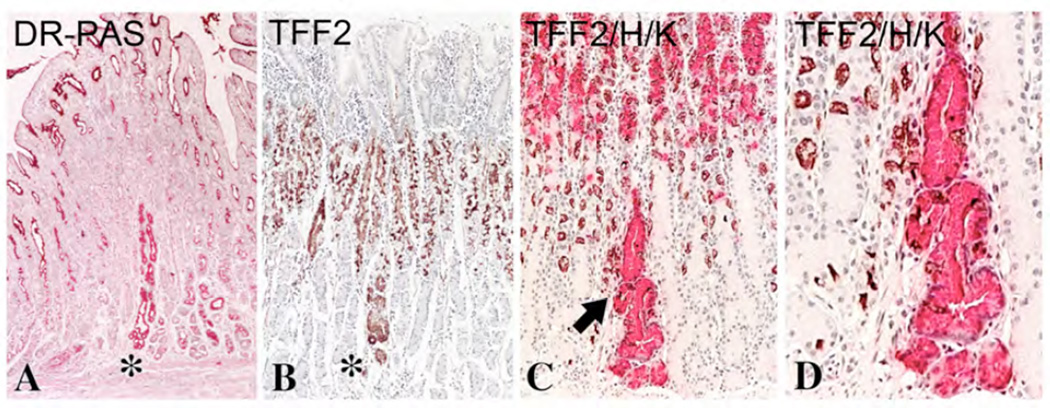
A. Diastase resistant-PAS (DR-PAS) staining of a section of human fundic mucosa showing the focal development of SPEM in a single gland unit (star). Note that in comparison with the carmine staining of surface cells, SPEM staining with DR-PAS is characteristically more reddish pink. B. TFF2 immunostaining staining with horseradish peroxidase secondary antibody staining and DAB (brown) chromagen showing a single gland unit with SPEM (star) surrounded by normal glands with TFF2-staining of mucous neck cells. C and D. Dual staining for TFF2 (red, alkaline phosphatase secondary antibody and Vector Red chromagen) and H/K-ATPase (brown DAB staining) staining of parietal cells showing the presence of one SPEM gland in the fundic mucosa.
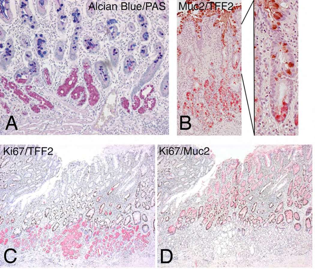
A. Dual Alcian Blue and PAS staining of a human fundic specimen showing compound glands with Alcian Blue staining intestinal metaplasia in more luminal cells and PAS-staining SPEM at the bases of glands. B. Dual Muc2 (brown) and TFF2 (red) immunostaining of a section of fundic mucosa showing glands with Muc2-immunoreactive intestinal metaplasia surmounting TFF2-staining SPEM. C and D. Serial sections of fundic mucosa from a resection specimen showing in C dual immunostaining for Ki67 (brown nuclei) and TFF2 (red) and in D dual immunostaining for Ki67 and Muc2 (red). While Ki67 staining nuclei can be seen in scattered SPEM cells, the majority of Ki67-staining cells nuclei are seen in Muc2-immunoreactive cells at the interface between SPEM and intestinal metaplasia.
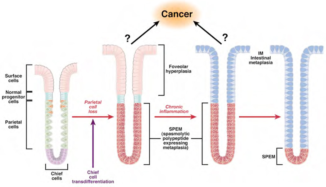
Loss of parietal cells leads to evolution of SPEM at the bases of glands from transdifferentiation of chief cells. With continuing chronic inflammation, intestinal metaplasia develops within the luminal aspect of SPEM glands. Over time, intestinal metaplasia comes to dominate over SPEM in metaplastic mucosa. In remains to be determined whether gastric cancer arises form SPEM or from proliferative intermediates generated during the further differentiation of SPEM into intestinal metaplasia.
References
-
- Correa P. A human model of gastric carcinogenesis. Cancer Res. 1988;48:3554–3560. - PubMed
-
- Zivny J, Wang TC, Yantiss R, Kim KH, Houghton J. Role of therapy or monitoring in preventing progression to gastric cancer. J Clin Gastroenterol. 2003;36:S50–60. discussion S61–52. - PubMed
-
- Schmidt PH, Lee JR, Joshi V, Playford RJ, Poulsom R, Wright NA, Goldenring JR. Identification of a metaplastic cell lineage associated with human gastric adenocarcinoma. Lab.Invest. 1999;79:639–646. - PubMed
-
- El-Zimaity HMT, Ota H, Graham DY, Akamatsu T, Katsuyama T. Patterns of gastric atrophy in intestinal type gastric carcinoma. Cancer. 2002;94:1428–1436. - PubMed
Publication types
MeSH terms
Substances
Grants and funding
LinkOut - more resources
Full Text Sources
Other Literature Sources
Medical
