. 2010 May 14;141(4):668-81.
doi: 10.1016/j.cell.2010.04.018. Epub 2010 May 6.

Peroxisomes are signaling platforms for antiviral innate immunity

Affiliations

Peroxisomes are signaling platforms for antiviral innate immunity

Evelyn Dixit et al. Cell. .

Abstract

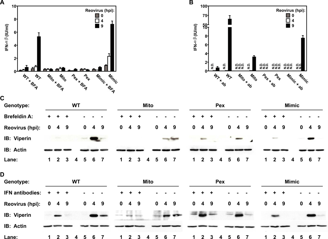
Peroxisomes have long been established to play a central role in regulating various metabolic activities in mammalian cells. These organelles act in concert with mitochondria to control the metabolism of lipids and reactive oxygen species. However, while mitochondria have emerged as an important site of antiviral signal transduction, a role for peroxisomes in immune defense is unknown. Here, we report that the RIG-I-like receptor (RLR) adaptor protein MAVS is located on peroxisomes and mitochondria. We find that peroxisomal and mitochondrial MAVS act sequentially to create an antiviral cellular state. Upon viral infection, peroxisomal MAVS induces the rapid interferon-independent expression of defense factors that provide short-term protection, whereas mitochondrial MAVS activates an interferon-dependent signaling pathway with delayed kinetics, which amplifies and stabilizes the antiviral response. The interferon regulatory factor IRF1 plays a crucial role in regulating MAVS-dependent signaling from peroxisomes. These results establish that peroxisomes are an important site of antiviral signal transduction.


Figures

Figure 1
Figure 1. MAVS resides on mitochondria and peroxisomes
(A) MEFs were transfected with the peroxisomal marker DsRed-PTS1 and either Flag-MAVS, myc-MFF or Flag-TIRAP. Cells were stained using anti-MAVS, anti-myc or anti-Flag antibodies, respectively. All images for all panels are representative of at least three independent experiments where over 500 cells were examined per condition and >95% of the cells displayed similar staining. (B) MEFs expressing Flag-MAVS as well as EGFP-PTS1 and Pex19 from a bicistronic construct were stained with anti-MAVS antibody and MitoTracker to visualize mitochondria. (C) Huh-7 hepatocytes were transfected with DsRed-PTS1 and endogenous MAVS was detected using anti-MAVS antisera. (D) Peroxisomes were separated from mitochondria on a Nycodenz gradient using HepG2 hepatocyte lysates. Selected fractions of the gradient were analyzed by immunoblotting with Pex14, mtHSP70, Fis1 or MAVS antisera. (E) Pex19-deficient human fibroblasts were stained for endogenous MAVS before and after introduction of a functional Pex19 allele as indicated. Mitochondria were stained with anti-mtHSP70 antibody. Peroxisomes were visualized by transfection with a bicistronic construct encoding EGFP-PTS1 and Pex19. See also Figure S1.
Figure 2
Figure 2. Targeting of MAVS to distinct subcellular compartments by replacement of its transmembrane domain
(A) Schematic of WT and mutant MAVS alleles to be tested for signaling from peroxisomes and mitochondria. (B) Stable cell lines expressing the MAVS alleles listed in (A) were generated by retroviral transduction of MAVS-KO cells. Resulting transgenic cells expressed a MAVS allele and GFP, whose translation is directed by an IRES. Shown are overlaid histograms of stable populations of each cell expressing equivalent levels of the bicistronic mRNAs encoding MAVS and GFP. (C) Lysates from stable cell lines described in (B) and parental MAVS KO MEFs were analyzed by immunoblotting with anti-MAVS antibody. (D) Micrographs of MAVS chimeric cell lines indicated were stained with anti-MAVS antibody. Mitochondria were stained with anti-mtHSP70 antibody. Peroxisomes were visualized by transfection with DsRed-PTS1. Note that MAVS-Pex resides on peroxisomes, MAVS-Mito on mitochondria, MAVS-Mimic on both organelles and MAVS-Cyto localizes on neither of the two organelles. All images for all panels are representative of at least three independent experiments where over 500 cells were examined per condition and >95% of the cells displayed similar staining. See also Figure S2.
Figure 3
Figure 3. Peroxisomal MAVS mediates ISG expression, but does not induce Type I IFN secretion
(A) MAVS-expressing MEFs and MAVS-KO cells were infected with reovirus. At indicated times, cell-associated ISG expression was determined by immunoblotting with an anti-viperin antibody. (B) Similar to (A) except for infection with influenza virus strain ΔNS1 in lieu of reovirus. (C and D) Cell culture media from (A) and (B) were tested for Type I IFN activity using a bioassay. Error bars show standard deviation of triplicate infections (E) MAVS-expressing MEFs and parental MAVS-KO cells were treated with 100 IU/ml IFNβ. At indicated times, cell associated ISG expression was determined by immunoblotting with anti-viperin antibody. Note that all cell lines respond similarly to IFNβ, indicating intact Type I IFN signaling. All data are the result of at least 2 independent experiments. See also Figure S3.
Figure 4
Figure 4. Peroxisomal MAVS directly induces viperin expression
(A) MAVS-expressing MEFs and MAVS-KO cells were pretreated with 20µg/ml BFA before infection with reovirus in presence of the drug. At indicated times, cell supernatants were tested for Type I IFN activity using a bioassay. (B) Similar to (A) except Type I IFN activity was blocked by addition of 250 NU/ml anti-IFNβ and 500 NU/ml anti-IFNα antibodies after infection with reovirus. (C and D) Cell lysates from (A) and (B) were tested for ISG expression by immunoblotting with anti-viperin antibody. Note that IFN activity is not required for viperin expression mediated by peroxisomal MAVS. All data are the representative of at least 3 independent experiments. Error bars show standard deviation of triplicate infections
Figure 5
Figure 5. Genome-wide transcriptome analysis reveals a general role for peroxisomal and mitochondrial MAVS in antiviral gene expression
(A) RNA from MAVS-expressing MEFs and parental MAVS-KO cells after infection with reovirus for 3, 9 or 16 h was subject to microarray analysis. The similarity of the overall gene expression profiles mediated by the indicated MAVS alleles is displayed as Pearson correlation coefficient-based heat map. Samples are clustered along both axes based on their correlation value. Note that at 3 h post infection MAVS-Pex cells display a gene expression pattern that is most similar to MAVS-WT cells. (B) Pairwise comparisons of indicated cell lines based on 4089 significantly regulated genes depicted in a log-log scale scatter plot. Each data point indicates a gene whose expression level exhibited a change of greater than 2-fold. (C) Heat map of selected genes based on their expression ratios across all 6 cell lines and during all time points upon reovirus infection. Genes are colored according to a log2-based color bar depicted underneath each heat map. See also Figure S4.
Figure 6
Figure 6. Peroxisomal MAVS elicits a functional antiviral response
(A) MAVS-expressing MEFs and MAVS-KO MEFs were infected with reovirus at an MOI of 3. At indicated times virus titers were determined by plaque assay. (B) MAVS-WT, -Mimic expressing cells and MAVS-KO MEFs were infected with VSV at an MOI of 3. After 8h RNA was isolated and analyzed for ISG and Type I IFN expression using nCounter. (C) Same as (B) except MAVS-Pex, MAVS-Mito and MAVS-Cyto MEFs were analyzed. (D) MAVS-expressing MEFs and parental MAVS-KO cells were infected with VSV at an MOI of 0.01. At indicated times, virus titers were determined by plaque assay. (E) 293T cells were transiently transfected with an ISRE, IRF1, NF-κB or AP1 luciferase reporters together with empty pMSCV vector (control) or MAVS (WT, Mimic, Pex, Mito or Cyto). Results were normalized to Renilla luciferase activity and are shown as fold increase relative to cells transfected with empty vector. Error bars show standard deviation of triplicate transfections. See also Figure S5.
Figure 7
Figure 7. Transcription factors that control peroxisomal MAVS signaling
(A,B) WT, IRF1, IRF3 and IRF5 KO MEFs were infected with VSV at an MOI of 3. After 8h RNA was isolated and analyzed for ISG and Type I IFN expression using nCounter. (C,D) Immortalized MAVS-KO macrophages were retrovirally transduced with MAVS-WT, -Pex, -Mito and-Cyto for 48 h. The transduction efficiency of each cell population was determined to be 20%–30% as assessed by fluorescence microscopy. Cells were infected with reovirus and at the indicated times, were harvested and RNA was analyzed for expression of Type I IFN (C) and other inflammatory genes (D) using nCounter. See also Figure S5. (E) Model of organelle specific MAVS signaling in fibroblasts. Peroxisomal MAVS is essential for rapid ISG expression independent of Type I IFN, whereas mitochondrial MAVS induces ISGs with delayed kinetics and primarily dependent on Type I IFN secretion. Therefore peroxisomal MAVS mediates immediate and transient antiviral effects, while mitochondrial MAVS promotes a sustained response later during infection. See also Figure S5.

Comment in

Similar articles

Cited by

References

    1. Ablasser A, Bauernfeind F, Hartmann G, Latz E, Fitzgerald KA, Hornung V. RIG-I-dependent sensing of poly(dA:dT) through the induction of an RNA polymerase III-transcribed RNA intermediate. Nat Immunol. 2009;10:1065–1072. - PMC - PubMed
    1. Akira S, Uematsu S, Takeuchi O. Pathogen recognition and innate immunity. Cell. 2006;124:783–801. - PubMed
    1. Amit I, Garber M, Chevrier N, Leite AP, Donner Y, Eisenhaure T, Guttman M, Grenier JK, Li W, Zuk O, et al. Unbiased reconstruction of a mammalian transcriptional network mediating pathogen responses. Science. 2009;326:257–263. - PMC - PubMed
    1. Balachandran S, Venkataraman T, Fisher PB, Barber GN. Fas-associated death domain-containing protein-mediated antiviral innate immune signaling involves the regulation of Irf7. J Immunol. 2007;178:2429–2439. - PubMed
    1. Barton GM, Kagan JC. A cell biological view of Toll-like receptor function: regulation through compartmentalization. Nat Rev Immunol. 2009;9:535–542. - PMC - PubMed

Publication types

Associated data