. 2010 Jun;47(6):500-6.
doi: 10.1016/j.ceca.2010.04.003. Epub 2010 May 6.

Skeletal muscle excitation-contraction coupling is independent of a conserved heptad repeat motif in the C-terminus of the DHPRbeta(1a) subunit

Affiliations

Skeletal muscle excitation-contraction coupling is independent of a conserved heptad repeat motif in the C-terminus of the DHPRbeta(1a) subunit

Anamika Dayal et al. Cell Calcium. 2010 Jun.

Abstract

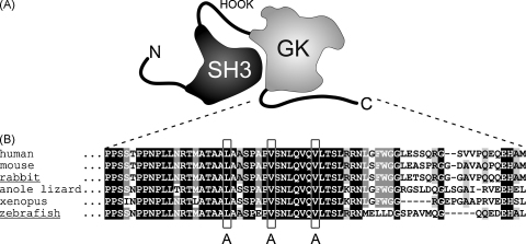
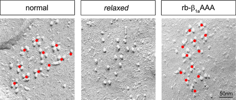
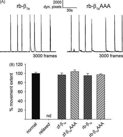
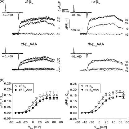
In skeletal muscle excitation-contraction (EC) coupling the sarcolemmal L-type Ca(2+) channel or 1,4-dihydropyridine receptor (DHPR) transduces the membrane depolarization signal to the sarcoplasmic Ca(2+) release channel RyR1 via protein-protein interaction. While it is evident that the pore-forming and voltage-sensing DHPRalpha(1S) subunit is essential for this process, the intracellular DHPRbeta(1a) subunit was also shown to be indispensable. We previously found that the beta(1a) subunit is essential to target the DHPR into groups of four (tetrads) opposite the RyR1 homotetramers, a prerequisite for skeletal muscle EC coupling. Earlier, a unique hydrophobic heptad repeat motif (Lcdots, three dots, centeredVcdots, three dots, centeredV) in the C-terminus of beta(1a) was postulated by others to be essential for skeletal muscle EC coupling, as substitution of these residues with alanines resulted in 80% reduction of RyR1 Ca(2+) release. Therefore, we wanted to address the question if the proposed beta(1a) heptad repeat motif could be an active element of the DHPR-RyR1 signal transduction mechanism or already contributes at the ultrastructural level i.e. DHPR tetrad arrangement. Surprisingly, our experiments revealed full tetrad formation and an almost complete restoration of EC coupling in beta(1)-null zebrafish relaxed larvae and isolated myotubes upon expression of a beta(1a)-specific heptad repeat mutant (LVV to AAA) and thus contradict the earlier results.


Figures

Fig. 1
Fig. 1
A conserved leucine-valine heptad repeat motif in the DHPRβ1a C-terminus. (A) Cartoon of the domain organization of the β1a subunit based on crystal structure models [28–30]. (B) Sequence alignment of β1a C-termini from different vertebrate classes, from fish to human, showed the conservation of the β1a-specific leucine-valine heptad repeat motif (boxed). To test for the contribution of the heptad repeat motif in skeletal muscle EC coupling, the LVV residues (boxed) were substituted by AAA (β1aAAA) in zebrafish and rabbit β1a subunits to be expressed in the zebrafish β1-null mutant relaxed system. Sequence for human (Homo sapiens) β1a, anole lizard (Anolis carolinensis) β1a, and Xenopus (Xenopus tropicalis) β1a were extracted from genomic assemblies at http://www.ensembl.org using a BLAST search with zf-β1a or rb-β1a cDNA sequences. GeneBank accession numbers for all other sequences used are: mouse (Mus musculus) β1a, NM_031173; rabbit (Oryctolagus cuniculus) β1a, NM_001082279 and zebrafish (Danio rerio) β1a, AY952462.
Fig. 2
Fig. 2
Intact DHPR tetrad formation with β1aAAA mutant expressed in relaxed myotubes. Freeze-fracture replicas of tail muscle tissue of normal zebrafish larvae (left panel) revealed the arrangement of DHPR particles in tetrads (indicated by red dots) organized in orthogonal arrays. In contrast, the β1-null mutant relaxed (middle panel) lacks DHPR tetrad formation. Comparable to normal larvae, relaxed larvae zygote-injected with in vitro synthesized rb-β1aAAA RNA, displayed correct assembly of DHPR particles in tetradic arrays (right panel). (For interpretation of the references to color in this figure legend, the reader is referred to the web version of the article.)
Fig. 3
Fig. 3
Full restoration of motility in relaxed larvae zygote-injected with β1aAAA mutant RNA. (A) Representative plots of total dynamic pixels per frame of 2-min video recordings of spontaneous larval movements. Relaxed larvae zygote-injected with either rb-β1a (left) or rb-β1aAAA RNA (right) revealed a similar movement profile. (B) To quantify larval movement extent, mean values of cumulative dynamic pixels per movement for each experimental group were calculated and standardized to those of normal larvae. Relaxed larvae zygote-injected with either zf-β1aAAA or rb-β1aAAA mutant RNA showed full recovery of larval movement extent, indistinguishable (p > 0.05) from normal larvae or from relaxed larvae injected with either zf-β1a or rb-β1a. One-way ANOVA revealed overall non-significance (p = 0.62, F(4,274) = 0.66). Uninjected relaxed larvae never showed any motility (nd, not detectable).
Fig. 4
Fig. 4
The β1aAAA mutation has only a minor effect on intracellular Ca2+ transients. (A) Representative intracellular Fluo-4 Ca2+ recordings from relaxed myotubes expressing zebrafish and rabbit WT β1a (upper panel) or β1aAAA mutant subunits (lower panel). Pronounced intracellular Ca2+ transients in response to 200-ms test pulses with similar kinetics were recorded with all constructs. Differences in transient size with β1aAAA can be explained by slight differences in expression levels as indicated by differences in intramembrane charge movement recordings (insets). (B) Voltage dependence curves of ΔF/F0 were corrected for differences in expression levels by normalizing ΔF/F0 to Qmax. Transients were slightly shifted towards more positive potentials for the β1aAAA constructs.

Similar articles

Cited by

References

    1. Schneider M.F., Chandler W.K. Voltage dependent charge movement of skeletal muscle: a possible step in excitation–contraction coupling. Nature. 1973;242:244–246. - PubMed
    1. Rios E., Brum G. Involvement of dihydropyridine receptors in excitation–contraction coupling in skeletal muscle. Nature. 1987;325:717–720. - PubMed
    1. Armstrong C.M., Bezanilla F.M., Horowicz P. Twitches in the presence of ethylene glycol bis(-aminoethyl ether)-N,N′-tetracetic acid. Biochim. Biophys. Acta. 1972;267:605–608. - PubMed
    1. Flucher B.E., Andrews S.B., Fleischer S., Marks A.R., Caswell A., Powell J.A. Triad formation: organization and function of the sarcoplasmic reticulum calcium release channel and triadin in normal and dysgenic muscle in vitro. J. Cell Biol. 1993;123:1161–1174. - PMC - PubMed
    1. Sun X.H., Protasi F., Takahashi M., Takeshima H., Ferguson D.G., Franzini-Armstrong C. Molecular architecture of membranes involved in excitation–contraction coupling of cardiac muscle. J. Cell Biol. 1995;129:659–671. - PMC - PubMed

Publication types

MeSH terms

Substances