. 2010 May 25;20(10):957-63.
doi: 10.1016/j.cub.2010.04.018. Epub 2010 May 6.

Systematic reduction of cohesin differentially affects chromosome segregation, condensation, and DNA repair

Affiliations

Systematic reduction of cohesin differentially affects chromosome segregation, condensation, and DNA repair

Jill M Heidinger-Pauli et al. Curr Biol. .

Abstract

Cohesin's complex distribution on chromosomes and its implication in numerous cellular processes makes it an excellent paradigm for studying the relationship between the in vivo concentration of a protein and its in vivo function. Here, we report a method to generate systematic quantized reductions (QR) in the in vivo concentration of any yeast protein. With QR, we generate strains with 13% and 30% of wild-type levels of the limiting subunit of cohesin, Mcd1p/Scc1p/Rad21p. Reducing cohesin levels reveals a preferential binding of cohesin to pericentric regions over cohesin-associated regions (CAR) on chromosome arms. Chromosome condensation, repetitive DNA stability, and DNA repair are compromised by decreasing cohesin levels to 30% of wild-type levels. In contrast, sister-chromatid cohesion and chromosome segregation are unaffected even when cohesin levels are reduced to 13% of wild-type levels. The requirement for different in vivo cohesin concentrations to achieve distinct cohesin functions provides an explanation for how cohesin mutations can specifically lead to adult disorders such as Cornelia de Lange Syndrome and Roberts Syndrome without compromising the cell divisions needed for development and maturation. Our successful application of QR to cohesin suggests that QR is a powerful tool to study other proteins/pathways with multiple functions.


Figures

Figure 1
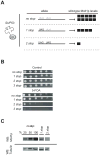
Figure 1. A method to gradually reduce cohesin levels in the cell demonstrates that the majority of cohesin is dispensable for cell viability
A. A stop codon is inserted in place of one or more Leucine (Leu) codons in the MCD1 ORF. Strains contain the SUP53 amber mutation, such that approximately 30% of the time Leu is inserted in place of each stop codon, resulting in the production of full length wild-type Mcd1p at a reduced frequency. B. JH2315-101 #15 was transformed with a plasmid containing CEN, LEU2 and mcd1 with the indicated number of stop mutations (pOM2–14 (no stop), pOM3–7 (1 stop), pNA1 (2 stop) and pCD1 (3 stop)). Transformants were grown in -LEU media overnight (30ºC) and 5X serial dilutions were plated on either -Leu plates (control) or 5-Fluoroorotic Acid (5-FOA) plates. No growth on 5-FOA represents an obligatory requirement for the wild-type MCD1 plasmid. C. JH5275a, JH5276b, JH5277b (0,1, 2 stop respectively integrated at the leu2 locus as the sole source of Mcd1p) were grown in YEPD to log phase at 30ºC. Cells were then arrested in G2/M using nocodazole (15 μg/ml final). Cells were pelleted, washed with dH2O, and frozen in liquid nitrogen. Pellets were processed for Sodium-polyacrylamide gel electrophoresis (SDS-PAGE) and Western blotting as described in experimental procedures. Quantification of bands (ratio of Mcd1p:Tub2p) in this experiment and a duplicate experiment using the IP labs software showed that the 1 stop contains approximately 30%(+/− 6.5%) while the 2 stop contains 13% (+/− 1.4%) of wild-type. N=2, a representative experiment is shown. See also Figure S1 for additional characterization of the growth of the QR strains.
Figure 2
Figure 2. Chromatin binding of QR cohesin strains
A. JH5275b, JH5276b, JH5277b (0, 1, 2 stop, respectively) were grown in YEPD to log phase at 30ºC. Cells were then arrested in G2/M using nocodazole (15μg/ml final). Cells were collected and processed for chromatin spreads (see experimental procedures), and Mcd1p was visualized using rabbit αMcd1p antibody. A representative field is shown from a representative experiment. N=3. B. Binding of cohesin along chromosome III as determined by chromatin immunoprecipitation. JH5275a, JH5276b, JH5277b (0,1, 2 stop, respectively) were grown in YEPD to log phase at 30ºC. Cells were then arrested in G2/M using nocodazole (15μg/ml final). Cells were fixed, collected and processed for chromatin immunoprecipitation (see experimental procedures) using rabbit α Mcd1p. The average cohesin binding from two independent experiments is plotted. C. Cohesin binding at three additional cohesin sites on Chr XIV and Chr XII. For binding of cohesin within the rDNA repeats, only 1.5μl of samples were used for the pcrs (instead of 2.5–5μl) and only 21 cycles were run (instead of 26) because of the large number of rDNA repeats. The binding we observe on chromosomes in the stop mutants is significant. Under conditions where cohesin is inactivated, Mcd1p binding drops, on average, to <0.2%. The fold changes in cohesin binding at the sites presented in this figure, averaged from two independent experiments, are presented in Figure S2.
Figure 3
Figure 3. Chromosome condensation, but not cohesion is affected in QR strains
JH5275a, JH5276b, JH5277b (0, 1, 2 stop, respectively) were grown in YEPD to log phase at 30ºC and then arrested in G2/M using nocodazole (15μg/ml final). Cells were fixed, collected and processed for fluorescent in situ hybridization (see experimental procedures) with a probe corresponding to 23–40kb from CEN16 (71042), 400kb away from CEN16 (70912), or against the rDNA repeats (pVG303). A total of 400 cells were counted for each experimental condition, from two independent experiments, and error bars indicate standard deviation. B. Images of a representative field from C. Green represents the rDNA probe, while DNA is stained with DAPI in red. C. Measurement distributions of the condensed state of the rDNA in JH5275a, JH5276b, and JH5277b. The size of the loop was measured as shown in the upper right hand corner of C. A single representative experiment is shown, where the number of cells counted =161 (no stop), 178 (1 stop) and 130 (2 stop).
Figure 4
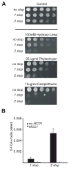
Figure 4. Cells with lower cohesin levels show increased marker loss from the rDNA and are sensitive to DSB-inducing drugs
A. JH5275b (no stop), JH5276b (1 stop), JH5277b (2 stop) were grown to saturation in YEPD, and then 5 fold serial dilutions were plated onto YEPD, 100mM Hydroxy-Urea, 0.1% MMS, .05μg/mL Phleomycin or 15μg/ml Camptothecin which were then incubated at 30ºC. Cell sensitivity to Benomyl is shown in Figure S3. B. JH5336 (1 stop) and JH5338 (2 stop) with or without plasmid pVG164(MCD1, TRP1, CEN) were grown on either –URA or –URA-TRP to select for URA3 integration at the rDNA and plasmid pVG164 respectively. Cells were picked up off of the plates and resuspended into dH2O and plated on 5-FOA or 5-FOA, -TRP or diluted and plated on YEPD plates to calculate the total number of cells plated.

Similar articles

Cited by

References

    1. Onn I, Heidinger-Pauli JM, Guacci V, Unal E, Koshland DE. Sister chromatid cohesion: a simple concept with a complex reality. Annu Rev Cell Dev Biol. 2008;24:105–129. - PubMed
    1. Liebman SW, Srodulski Z, Reed CR, Stewart JW, Sherman F, Brennan G. Yeast amber suppressors corresponding to tRNA3Leu genes. J Mol Biol. 1984;178:209–226. - PubMed
    1. Little JW. Mutants of bacteriophage T4 which allow amber mutants of gene 32 to grow in ochre-suppressing hosts. Virology. 1973;53:47–59. - PubMed
    1. Michaelis C, Ciosk R, Nasmyth K. Cohesins: chromosomal proteins that prevent premature separation of sister chromatids. Cell. 1997;91:35–45. - PubMed
    1. Toth A, Ciosk R, Uhlmann F, Galova M, Schleiffer A, Nasmyth K. Yeast cohesin complex requires a conserved protein, Eco1p(Ctf7), to establish cohesion between sister chromatids during DNA replication. Genes Dev. 1999;13:320–333. - PMC - PubMed

Publication types

MeSH terms

Substances