. 2010 May 25;20(10):887-94.
doi: 10.1016/j.cub.2010.03.066. Epub 2010 May 6.

Perceptual learning improves contrast sensitivity of V1 neurons in cats

Affiliations

Perceptual learning improves contrast sensitivity of V1 neurons in cats

Tianmiao Hua et al. Curr Biol. .

Abstract

Background: Perceptual learning has been documented in adult humans over a wide range of tasks. Although the often-observed specificity of learning is generally interpreted as evidence for training-induced plasticity in early cortical areas, physiological evidence for training-induced changes in early visual cortical areas is modest, despite reports of learning-induced changes of cortical activities in fMRI studies. To reveal the physiological bases of perceptual learning, we combined psychophysical measurements with extracellular single-unit recording under anesthetized preparations and examined the effects of training in grating orientation identification on both perceptual and neuronal contrast sensitivity functions of cats.

Results: We have found that training significantly improved perceptual contrast sensitivity of the cats to gratings with spatial frequencies near the "trained" spatial frequency, with stronger effects in the trained eye. Consistent with behavioral assessments, the mean contrast sensitivity of neurons recorded from V1 of the trained cats was significantly higher than that of neurons recorded from the untrained cats. Furthermore, in the trained cats, the contrast sensitivity of V1 neurons responding preferentially to stimuli presented via the trained eyes was significantly greater than that of neurons responding preferentially to stimuli presented via the "untrained" eyes. The effect was confined to the trained spatial frequencies. In both trained and untrained cats, the neuronal contrast sensitivity functions derived from the contrast sensitivity of the individual neurons were highly correlated with behaviorally determined perceptual contrast sensitivity functions.

Conclusions: We suggest that training-induced neuronal contrast gain in area V1 underlies behaviorally determined perceptual contrast sensitivity improvements.


Figures

Fig. 1
Fig. 1
Visual stimuli used in conditioning training of cat1 (A; 0.2 c/deg) and cat2 (B; 0.4 c/deg). All stimuli are 80% contrast sine wave gratings oriented at ±45 degrees.
Fig. 2
Fig. 2
Contrast sensitivity functions in the trained and untrained eyes before and after training for cat1 (A) and cat2 (B). Smooth curves represent the best fitting Gauss functions. The green arrows indicate the trained spatial frequency, and the error bars represent 1 SD.
Fig. 3
Fig. 3
Contrast sensitivity at the trained spatial frequency (expressed as mean ± SD) versus training days for cat1 (filled circles) and cat2 (open circles).
Fig. 4
Fig. 4
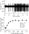
A typical neuron's response to its optimal visual stimulus. A: The voltage trace of the neuron's response to the optimal stimulus at 64% contrast. A spike with amplitude surpassing the horizontal broken line is counted as an action potential. The neuron's response is evoked by 5 cycles of grating stimulation, equivalent to a stimulus duration of about 1.7 seconds, and the spontaneous activity (M) is acquired 1 second prior to visual stimulus presentation. The arrowhead indicates the stimulus onset time. B: Contrast response function of the neuron (mean±SD). The smooth curve represents the best fitting Naka-Rushton equation (r2=99.5%). M and Rmax represent the neuron's spontaneous activity and maximal visually-evoked response to visual stimuli. TC (threshold stimulus contrast) represents the stimulus contrast that evokes a neuron's response that is 1.414 times of its spontaneous activity. C50 corresponds to the stimulus contrast that evokes half of the neuron's maximal response. N represents the slope of the neuron's response-contrast tuning curve.
Fig. 5
Fig. 5
(AB) TC-contrast sensitivity functions of V1 neurons recorded from cat1 (A) and cat2 (B). (CD) C50-contrast sensitivity functions of V1 neurons recorded from cat1 (C) and cat2 (D). Green arrows indicate the trained spatial frequency. All values are displayed as mean ± SEM.
Fig. 6
Fig. 6
Scatter plots of mean neuronal contrast sensitivity versus mean psychophysical contrast sensitivity across different spatial frequencies (0.1, 0.2, 0.4, 0.6, 0.8, 1.2 and 1.6 c/deg) for the trained and untrained eyes of the trained cats (red circle: trained eye of trained cat1; purple circle: untrained eye of trained cat1; blue circle: trained eye of trained cat2; green circle: untrained eye of trained cat2). Neuronal contrast sensitivity is based on TC in Panel I, and based on C50 in panel II. (A) and (B) contrast sensitivities before training. (C) and (D) contrast sensitivities after training. Colored lines in each subplot represent the best linear fits (Red: trained eye of trained cat1; Purple: untrained eye of trained cat1; Blue: trained eye of trained cat2; Green: untrained eye of trained cat2.).
Fig. 7
Fig. 7
Parameters of the best-fitting Naka-Rushton equation to the neuronal contrast response functions. (A) Spontaneous activities. (B) Maximum responses. (C) Slopes of the contrast response functions (SL). (D) Contrast-gain (C50) of cells in each trained and control cat. (E) Contrast-gain of cells with optimal SF 0.2-0.6 c/deg in trained cat1. (F) Contrast-gain of cells with optimal SF 0.4-0.8 c/deg in trained cat2. TE and NE denote cells responding preferentially to the stimuli presented via the trained eye and the naïve eye. All values are expressed as mean ± SEM.

References

    1. Fiorentini A, Berardi N. Perceptual learning specific for orientation and spatial frequency. Nature. 1980;287:43–44. - PubMed
    1. Kami A, Sagi D. Where practice makes perfect in texture discrimination: evidence for primary visual cortex plasticity. Proc Natl Acad Sci U S A. 1991;88:4966–4970. - PMC - PubMed
    1. Liu Z, Vaina LM. Simultaneous learning of motion discrimination in two directions. Cognitive Brain Research. 1998;6:347–349. - PubMed
    1. Sowden PT, Rose D, Davies IR. Perceptual learning of luminance contrast detection: specific for spatial frequency and retinal location but not orientation. Vision Res. 2002;42:1249–1258. - PubMed
    1. Gilbert CD. Early perceptual learning. Proc Natl Acad Sci U S A. 1994;91:1195–1197. - PMC - PubMed

Publication types