Review
. 2011 Feb;1808(2):522-9.
doi: 10.1016/j.bbamem.2010.04.015. Epub 2010 May 6.

Influenza M2 proton channels

Affiliations
Review

Influenza M2 proton channels

Rafal M Pielak et al. Biochim Biophys Acta. 2011 Feb.

Abstract

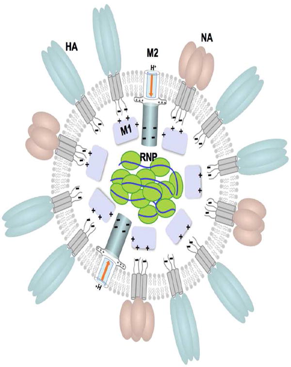
M2 of the influenza virus is an intriguing transmembrane protein that forms a minuscule proton channel in the viral envelope. Its recognized function is to equilibrate pH across the viral membrane during cell entry and across the trans-Golgi membrane of infected cells during viral maturation. It is vital for viral replication and it is a target for the anti-influenza drugs, amantadine and rimantadine. Recently, high resolution structures of M2 channels of both flu A and B have been obtained, providing the desperately needed structural details for understanding the mechanism of proton conductance. In particular, the establishment of the functional solution NMR system of the proton channels enabled simultaneous high resolution structure characterization and measurement of channel dynamics coupled to channel activity. This review summarizes our current understanding of how protons are conducted through the M2 channel from a structural point of view, as well as the modes by which important channel gating elements function during proton conduction.


Figures

Figure 1
Figure 1
Viral particle of influenza with its membrane proteins: hemagglutinin – HA, neuraminidase – NA, matrix protein 2 M2; and associated proteins: matrix protein 1 – M1, ribonucleoprotein – RNP (A) A model of the viral particle. (B) Viral budding: HA of influenza A (PDB code: 5HMG), NA of influenza A (PDB code: 1ING), M2 of influenza B (PDB code: 2KIV and 2KJ1).
Figure 1
Figure 1
Viral particle of influenza with its membrane proteins: hemagglutinin – HA, neuraminidase – NA, matrix protein 2 M2; and associated proteins: matrix protein 1 – M1, ribonucleoprotein – RNP (A) A model of the viral particle. (B) Viral budding: HA of influenza A (PDB code: 5HMG), NA of influenza A (PDB code: 1ING), M2 of influenza B (PDB code: 2KIV and 2KJ1).
Figure 2
Figure 2
High resolution structures of the AM2 channel domain. (A) Solution structure of residues 18 – 60 in DHPC (1,2-Dihexanoyl-sn-Glycero-3-Phosphocholine) micelles at pH 7.5 (PDB code: 2RLF) (Schnell and Chou 2008). (B) Crystal structure of residues 22 – 46 with the I33Se-Met mutation in OG (octyl-β-D-glucopyranoside) micelles at pH 7.3 (PDB code: 3BKD) (Stouffer, Acharya et al. 2008).
Figure 3
Figure 3
The BM2 structures. (A) Solution structures of BM2 channel (residues 1 – 33) and cytoplasmic (residues 44 – 103) domains. The channel domain (PDB code: 2KIX)(Wang, Pielak et al. 2009) is positioned relative to the hydrophobic region of the presumed lipid bilayer as defined by two lines. The structure of the linker region (residues 34 – 43) has not been defined. The cytoplasmic domain (PDB code: 2KJ1)(Wang, Pielak et al. 2009) interacts with the BM1 matrix protein, probably through the BM1 N-terminal domain shown on the right. (B) Helical wheel representation of the BM2(1-33) coiled coil tetramer. Polar resides are presented in red.
Figure 4
Figure 4
Structural elements of AM2 and BM2 important for channel function. (A) and (B) show the C-terminal tryptophan gates of the AM2 (PDB code: 2RLF)(Schnell and Chou 2008) and BM2 (PDB code: 2KIX)(Wang, Pielak et al. 2009) channels, respectively. (C) and (D) show the amino acid sidechains important for proton relay, selection, and gating in the TM domains of AM2 and BM2, respectively. (E) and (F) show the N-terminal constriction of the AM2 and BM2 channel, respectively.

Similar articles

Cited by

References

    1. Palese P, Shaw ML. Orthomyxoviridae: the viruses and their replication. In: Knipe DM, Howley PM, editors. Fields virology. Williams & Wilkins; 2007.
    1. Bouvier NM, Palese P. The biology of influenza viruses. Vaccine. 2008;26(Suppl 4):D49–53. - PMC - PubMed
    1. Zebedee SL, Lamb RA. Influenza A virus M2 protein: monoclonal antibody restriction of virus growth and detection of M2 in virions. J Virol. 1988;62:2762–2772. - PMC - PubMed
    1. Lakadamyali M, Rust MJ, Babcock HP, Zhuang X. Visualizing infection of individual influenza viruses. Proc Natl Acad Sci U S A. 2003;100:9280–9285. - PMC - PubMed
    1. Hay AJ, Wolstenholme AJ, Skehel JJ, Smith MH. The molecular basis of the specific anti-influenza action of amantadine. EMBO J. 1985;4:3021–3024. - PMC - PubMed

LinkOut - more resources