

In vivo voltammetric monitoring of catecholamine release in subterritories of the nucleus accumbens shell
- PMID: 20451589
- PMCID: PMC2900378
- DOI: 10.1016/j.neuroscience.2010.04.076
In vivo voltammetric monitoring of catecholamine release in subterritories of the nucleus accumbens shell
Abstract
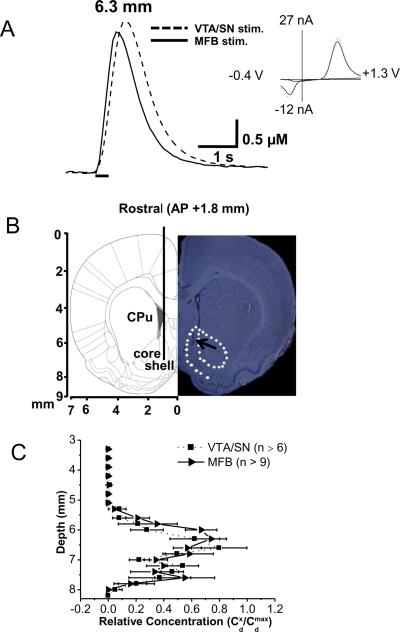
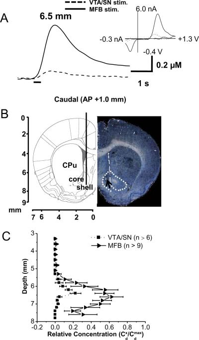
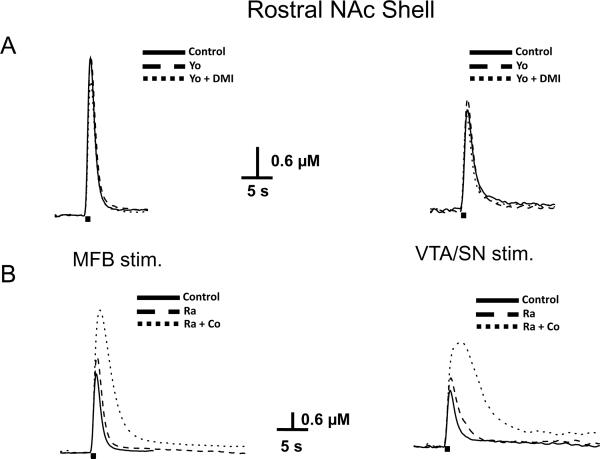
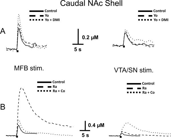
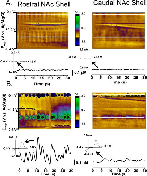
Fast-scan cyclic voltammetry (FSCV) at carbon-fiber microelectrodes has been used to demonstrate that sub-second changes in catecholamine concentration occur within the nucleus accumbens (NAc) shell during motivated behaviors, and these fluctuations have been attributed to rapid dopamine signaling. However, FSCV cannot distinguish between dopamine and norepinephrine, and caudal regions of the NAc shell receive noradrenergic projections. Therefore, in the present study, we examined the degree to which norepinephrine contributes to catecholamine release within rostral and caudal portion of NAc shell. Analysis of tissue content revealed that dopamine was the major catecholamine detectable in the rostral NAc shell, whereas both dopamine and norepinephrine were found in the caudal subregion. To examine releasable catecholamines, electrical stimulation was used to evoke release in anesthetized rats with either stimulation of the medial forebrain bundle, a pathway containing both dopaminergic and noradrenergic projections to the NAc, or the ventral tegmental area/substantia nigra, the origin of dopaminergic projections. The catecholamines were distinguished by their responses to different pharmacological agents. The dopamine autoreceptor blocker, raclopride, as well as the monoamine and dopamine transporter blockers, cocaine and GBR 12909, increased evoked catecholamine overflow in both the rostral and caudal NAc shell. The norepinephrine autoreceptor blocker, yohimbine, and the norepinephrine transporter blocker, desipramine, increased catecholamine overflow in the caudal NAc shell without significant alteration of evoked responses in the rostral NAc shell. Thus, the neurochemical and pharmacological results show that norepinephrine signaling is restricted to caudal portions of the NAc shell. Following raclopride and cocaine or raclopride and GBR 12909, robust catecholamine transients were observed within the rostral shell but these were far less apparent in the caudal NAc shell, and they did not occur following yohimbine and desipramine. Taken together, the data demonstrate that catecholamine signals in the rostral NAc shell detected by FSCV are due to change in dopamine transmission.
Copyright (c) 2010 IBRO. Published by Elsevier Ltd. All rights reserved.
Figures
References
-
- Baur JE, Kristensen EW, May LJ, Wiedemann DJ, Wightman RM. Fast-scan voltammetry of biogenic amines. Anal Chem. 1988;60:1268–1272. - PubMed
-
- Berridge CW, Stratford TL, Foote SL, Kelley AE. Distribution of dopamine beta-hydroxylase-like immunoreactive fibers within the shell subregion of the nucleus accumbens. Synapse. 1997;27:230–241. - PubMed
-
- Budygin EA, Kilpatrick MR, Gainetdinov RR, Wightman RM. Correlation between behavior and extracellular dopamine levels in rat striatum: comparison of microdialysis and fast-scan cyclic voltammetry. Neurosci Lett. 2000;281:9–12. - PubMed
Publication types
MeSH terms
Substances
Grants and funding
LinkOut - more resources
Full Text Sources
