. 2010 Sep;39(3):311-7.
doi: 10.1016/j.nbd.2010.04.015. Epub 2010 May 6.

Inhibition of c-Jun kinase provides neuroprotection in a model of Alzheimer's disease

Affiliations

Inhibition of c-Jun kinase provides neuroprotection in a model of Alzheimer's disease

Steven P Braithwaite et al. Neurobiol Dis. 2010 Sep.

Abstract

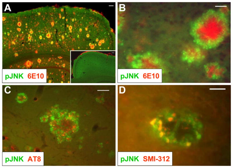
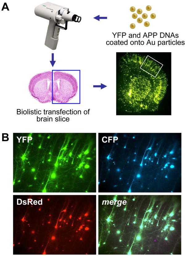
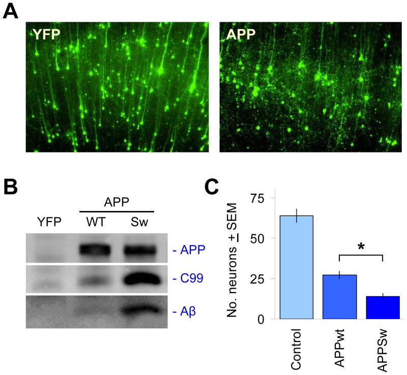
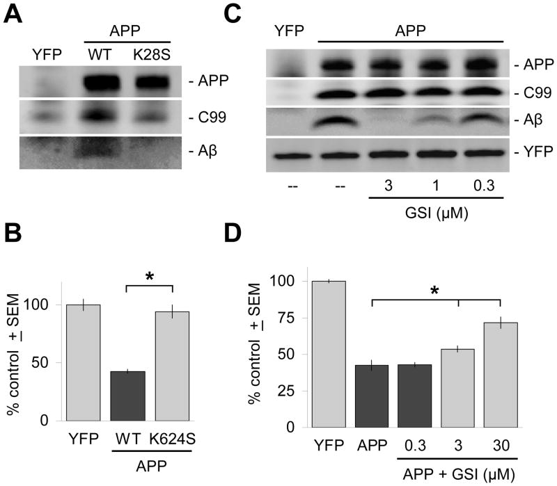
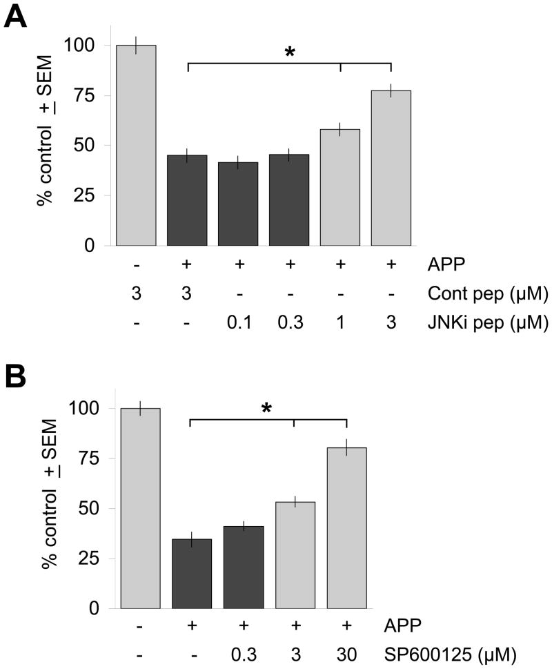
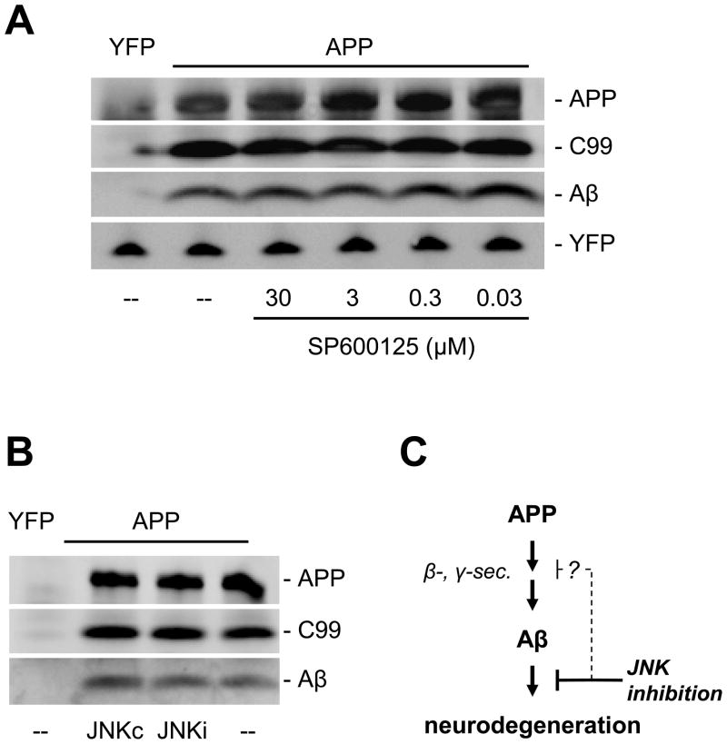
The c-Jun N-terminal kinase (JNK) pathway potentially links together the three major pathological hallmarks of Alzheimer's disease (AD): development of amyloid plaques, neurofibrillary tangles, and brain atrophy. As activation of the JNK pathway has been observed in amyloid models of AD in association with peri-plaque regions and neuritic dystrophy, as we confirm here for Tg2576/PS(M146L) transgenic mice, we directly tested whether JNK inhibition could provide neuroprotection in a novel brain slice model for amyloid precursor protein (APP)-induced neurodegeneration. We found that APP/amyloid beta (Abeta)-induced neurodegeneration is blocked by both small molecule and peptide inhibitors of JNK, and provide evidence that this neuroprotection occurs downstream of APP/Abeta production and processing. Our findings demonstrate that Abeta can induce neurodegeneration, at least in part, through the JNK pathway and suggest that inhibition of JNK may be of therapeutic utility in the treatment of AD.


Figures

Figure 1
Figure 1. JNK is activated in dystrophic neurons in the peri-plaque region in Tg2576/PS1M146L mice
(A) 12-month old Tg2576/PS1M146L transgenic mice show strong 6E10 immunoreactivity indicative of Aβ deposition (red) and increased JNK phosphorylation (green) in the cortex compared to wild-type mice (inset). Scale bar, 100 μm. (B) At higher magnification, phosphorylated JNK (green) is seen to surround amyloid plaques (red) in cortex. Scale bar, 20 μm. (C) JNK phosphorylation (green) partially overlaps with immunoreactivity with the AT8 antibody, indicative of hyperphosphorylated tau. Scale bar, 50 μm. (D) JNK phosphorylation (green) also coincides with SMI-312 immunoreactivity, indicative of dystrophic neurons. Scale bar, 20 μm. All images are representative of observations from at least 3 animals.
Figure 2
Figure 2. Biolistic transfection is used to introduce APP isoforms into organotypic brain slice explants
(A) 1.6 μm elemental gold particles coated with YFP and APP isoform DNA expression plasmids are accelerated with a gene gun to transfect P10 rat coronal brain slice explants, resulting in even transfection of resident neuronal cell types in the explants (lower right photomicrograph), including pyramidal neurons in the cortex (white boxed region). (B) Co-transfection linkage rate using biolistics is >99%. Co-coating gold particles with 3 different DNA plasmids each encoding a different fluorescent protein (YFP, CFP, and DsRed) leads to clear co-expression in essentially all transfected neurons shown here in a magnified portion of the cortex (merge at lower right).
Figure 3
Figure 3. Acute challenge with APP isoforms leads to its proteolytic processing into Aβ and the induction of neuronal degeneration
(A) The co-transfected YFP fluorescent marker clearly delineates neuronal cell bodies and dendrites after 2–3 d in culture, shown in the left panel for a region in cortex (pia surface up). It can be seen that the majority of neurons transfected are cortical pyramidal neurons, easily distinguished by their extension of a single, prominent apical dendrite in the radial direction. Right panel shows overt neurodegeneration of such cortical pyramidal neurons 2–3 days after co-transfection with APP isoforms such as APPSw. (B) Expression of APP is readily detected in transfected brain slices via immunoprecipitation and immunoblotting with the human-specific APP antibody 6E10. Processing of APP into C99 and Aβ by native tissue proteases can also be seen, with higher levels of C99 and Aβ production as expected for APPSw relative to APPWT. (C) Induction of neurodegeneration by transfected APP is more severe with the Swedish mutation. Ordinate axis shows average, total numbers ± SEM of non-degenerating pyramidal neurons in the cortical regions of each explant, N=12 brain slices scored per condition. *, significant by ANOVA followed by Dunnett’s post hoc comparison test at the 0.05 confidence level.
Figure 4
Figure 4. Inhibition of γ-secretase cleavage inhibits Aβ production and is neuroprotective
(A) The APPK624S mutation inhibits γ-secretase cleavage of APP and decreases Aβ production to undetectable levels in 6E10 immunoblots of brain slice lysates, but does not significantly affect levels of expression of full-length APP. (B) Correspondingly, transfection with the APPK624S mutation does not result in measurable levels of neurodegeneration. (C) Treatment of transfected brain slices with the γ-secretase inhibitor WYGSI-04 (“GSI”) for 2 days resulted in selective, dose-dependent inhibition of Aβ production. Such immunoblot analysis showed that WYGSI-04 treatment had no effect on the expression of full-length APP, or on its initial cleavage by endogenous β-secretases to the intermediate fragment C99. Lanes were loaded with identical amounts of total protein from brain slice lysates, with equivalent transfection rates confirmed by direct immunoblotting against the co-transfected marker YFP (bottom band). “--” denotes treatment with the DMSO vehicle only. (D) Correspondingly, inhibition of Aβ production by WYGSI-04 provides dose-dependent neuroprotection in the same concentration range. Ordinate axes in (B) and (D) show average, total numbers ± SEM of non-degenerating pyramidal neurons in the cortical regions of each explant expressed as a percentage of control brain slices transfected with YFP only. *, significant by ANOVA followed by Dunnett’s post hoc comparison test at the 0.05 confidence level.
Figure 5
Figure 5. Inhibition of JNK activity reduces neurodegeneration induced by APP/Aβ
(A) Treatment of APP-transfected brain slices with the cell-permeable JNK inhibitory peptide JNKi (“JNKi pep”) protected against APP/Aβ-induced neurodegeneration in a dose-dependent manner, while a control peptide containing only the HIV-TAT N-terminal domain (“Cont pep”) had no effect. (B) The small molecule JNK inhibitor SP600125 also provided dose-dependent rescue of neurodegeneration induced by APP transfection. Ordinate axes show average, total numbers ± SEM of non-degenerating pyramidal neurons in the cortical regions of each explant expressed as a percentage of control brain slices transfected with YFP only and treated as indicated. *, significant differences by ANOVA followed by Dunnett’s post hoc comparison test at the 0.05 confidence level.
Figure 6
Figure 6. JNK inhibition does not affect the production of Aβ
Incubation of transfected brain slices with concentrations of the JNK inhibitor SP600125 (A) or JNKi peptide (B) across the ranges that provided neuroprotection showed no changes in expression levels of full-length APP, C99, or Aβ. Lanes were loaded with identical amounts of total protein from brain slice lysates, with equivalent transfection rates confirmed by direct immunoblot against the co-transfected marker YFP (bottom band). “--” denotes treatment with the DMSO vehicle only. (C) Together, these findings suggest that the primary site of action of JNK inhibition in this brain slice model for APP/Aβ-induced neurodegeneration lies downstream of the production of Aβ. However, other studies have suggested that JNK may also play roles in the production of Aβ itself (Colombo et al., 2009).

References

    1. Barr RK, et al. Identification of the critical features of a small peptide inhibitor of JNK activity. J Biol Chem. 2002;277:10987–97. - PubMed
    1. Bennett BL, et al. SP600125, an anthrapyrazolone inhibitor of Jun N-terminal kinase. Proc Natl Acad Sci U S A. 2001;98:13681–6. - PMC - PubMed
    1. Borsello T, et al. A peptide inhibitor of c-Jun N-terminal kinase protects against excitotoxicity and cerebral ischemia. Nat Med. 2003;9:1180–6. - PubMed
    1. Brecht S, et al. Specific pathophysiological functions of JNK isoforms in the brain. Eur J Neurosci. 2005;21:363–77. - PubMed
    1. Cho S, et al. Brain Slices as Models for Neurodegenerative Disease and Screening Platforms to Identify Novel Therapeutics. Current Neuropharmacology. 2007;5:19–33. - PMC - PubMed

Publication types

MeSH terms

Substances