

Update on the magnetic resonance imaging core of the Alzheimer's disease neuroimaging initiative
- PMID: 20451869
- PMCID: PMC2886577
- DOI: 10.1016/j.jalz.2010.03.004
Update on the magnetic resonance imaging core of the Alzheimer's disease neuroimaging initiative
Abstract
Functions of the Alzheimer's Disease Neuroimaging Initiative (ADNI) magnetic resonance imaging (MRI) core fall into three categories: (1) those of the central MRI core laboratory at Mayo Clinic, Rochester, Minnesota, needed to generate high quality MRI data in all subjects at each time point; (2) those of the funded ADNI MRI core imaging analysis groups responsible for analyzing the MRI data; and (3) the joint function of the entire MRI core in designing and problem solving MR image acquisition, pre-processing, and analyses methods. The primary objective of ADNI was and continues to be improving methods for clinical trials in Alzheimer's disease. Our approach to the present ("ADNI-GO") and future ("ADNI-2," if funded) MRI protocol will be to maintain MRI methodological consistency in the previously enrolled "ADNI-1" subjects who are followed up longitudinally in ADNI-GO and ADNI-2. We will modernize and expand the MRI protocol for all newly enrolled ADNI-GO and ADNI-2 subjects. All newly enrolled subjects will be scanned at 3T with a core set of three sequence types: 3D T1-weighted volume, FLAIR, and a long TE gradient echo volumetric acquisition for micro hemorrhage detection. In addition to this core ADNI-GO and ADNI-2 protocol, we will perform vendor-specific pilot sub-studies of arterial spin-labeling perfusion, resting state functional connectivity, and diffusion tensor imaging. One of these sequences will be added to the core protocol on systems from each MRI vendor. These experimental sub-studies are designed to demonstrate the feasibility of acquiring useful data in a multicenter (but single vendor) setting for these three emerging MRI applications.
Copyright 2010 The Alzheimer
Figures
References
-
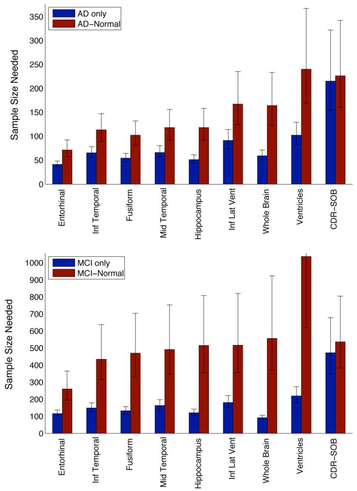
- Holland D, Brewer JB, Hagler DJ, Fenema-Notestine C, Dale AM, Weiner M, Thal L, Petersen R, Jack CR, Jr, Jagust W, Trojanowki J, Toga AW, Beckett L, Green RC, Gamst A, Potter WZ, Montine T, Anders D, Bernstein M, Felmlee J, Fox N, Thompson P, Schuff N, Alexander G, Bandy D, Koeppe RA, Foster N, Reiman EM, Chen K, Shaw L, Lee VM, Korecka M, Crawford K, Neu S, Harvey D, Kornak J, Kachaturian Z, Frank R, Snyder PJ, Molchan S, Kaye J, Vorobik R, Quinn J, Schneider L, Pawluczyk S, Spann B, Fleisher AS, Vanderswag H, Heidebrink JL, Lord JL, Johnson K, Doody RS, Villanueva-Meyer J, Chowdhury M, Stern Y, Honig LS, Bell KL, Morris JC, Mintun MA, Schneider S, Marson D, Griffith R, Badger B, Grossman H, Tang C, Stern J, Detoledo-Morrell L, Shah RC, Bach J, Duara R, Isaacson R, Strauman S, Albert MS, Pedroso J, Toroney J, Rusinek H, de Leon MJ, De Santi SM, Doraiswamy PM, Petrella JR, Aiello M, Clark CM, Pham C, Nunez J, Smith CD, Given CA, 2, Hardy P, Dekosky ST, Oakley M, Simpson DM, Ismail MS, Porsteinsson A, McCallum C, Cramer SC, Mulnard RA, McAdams-Ortiz C, Diaz-Arrastia R, Martin-Cook K, Devous M, Levey AI, Lah JJ, Cellar JS, Burns JM, Anderson HS, Laubinger MM, Bartzokis G, Silverman DH, Lu PH, Fletcher R, Parfitt F, Johnson H, Farlow M, Herring S, Hake AM, van Dyck CH, Macavoy MG, Bifano LA, Chertkow H, Bergman H, Hosein C, Black S, Graham S, Caldwell C, Feldman H, Assaly M, Hsiung GY, Kertesz A, Rogers J, Trost D, Bernick C, Gitelman D, Johnson N, Mesulam M, Sadowsky C, Villena T, Mesner S, Aisen PS, Johnson KB, Behan KE, Sperling RA, Rentz DM, Johnson KA, Rosen A, Tinklenberg J, Ashford W, Sabbagh M, Connor D, Obradov S, Killiany R, Norbash A, Obisesan TO, Jayam-Trouth A, Wang P, Auchus AP, Huang J, Friedland RP, Decarli C, Fletcher E, Carmichael O, Kittur S, Mirje S, Johnson SC, Borrie M, Lee TY, Asthana S, Carlsson CM, Potkin SG, Highum D, Preda A, Nguyen D, Tariot PN, Hendin BA, Scharre DW, Kataki M, Beversdorf DQ, Zimmerman EA, Celmins D, Brown AD, Gandy S, Marenberg ME, Rovner BW, Pearlson G, Blank K, Anderson K, Saykin AJ, Santulli RB, Pare N, Williamson JD, Sink KM, Potter H, Ashok Raj B, Giordano A, Ott BR, Wu CK, Cohen R, Wilks KL, Safirstein BE. Subregional neuroanatomical change as a biomarker for Alzheimer's disease. Proc Natl Acad Sci U S A. 2009 Dec 8;106(49):20954–9. - PMC - PubMed
-
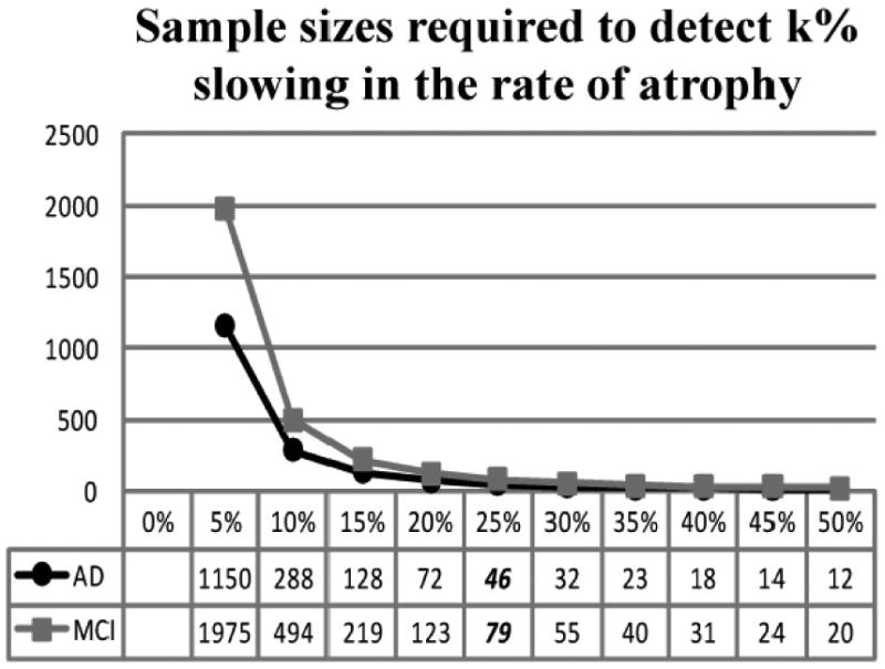
- Hua X, Lee S, Hibar DP, Yanovsky I, Leow AD, Toga AW, Jack CR, Jr, Bernstein MA, Reiman EM, Harvey DJ, Kornak J, Schuff N, Alexander GE, Weiner MW, Thompson PM. Mapping Alzheimer's disease progression in 1309 MRI scans: Power estimates for different inter-scan intervals. Neuroimage. Feb 6; - PMC - PubMed
-
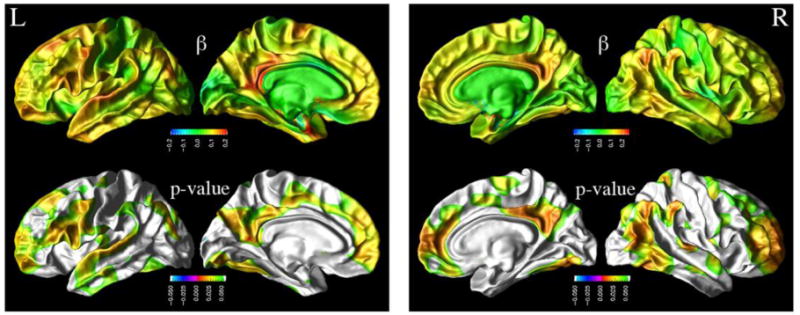
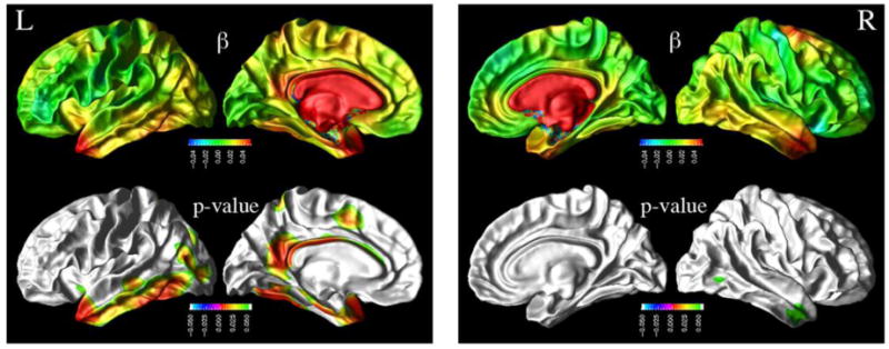
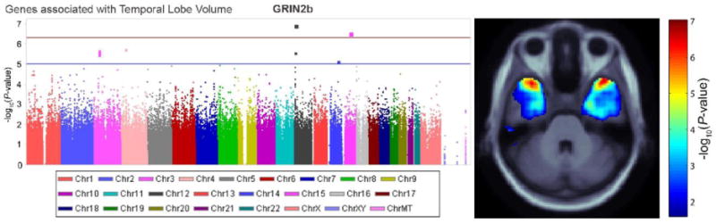
- Stein JL, Hua X, Morra JH, Lee S, Ho AJ, Leow AD, Toga AW, Sul J, Kang HM, Eskin E, Saykin AJ, Shen L, Foroud T, Pankratz N, Huentelman MJ, Craig DW, Gerber JD, Allen A, Corneveaux J, Stephan DA, Webster J, DeChairo BM, Potkin SG, Jack CR, Jr, Weiner MW. Genome-wide association study of temporal lobe structure identifies novel quantitative trait loci for neurodegeneration in alzheimer's disease. Neuron. 2009 Submitted July 21 2009.
-
- Freeborough PA, Fox NC. The boundary shift integral: an accurate and robust measure of cerebral volume changes from registered repeat MRI. IEEE Trans Med Imaging. 1997 Oct;16(5):623–9. - PubMed
Publication types
MeSH terms
Substances
Grants and funding
LinkOut - more resources
Full Text Sources
Medical
