. 2010 May;6(3):221-9.
doi: 10.1016/j.jalz.2010.03.003.

The Alzheimer's Disease Neuroimaging Initiative positron emission tomography core

Affiliations

The Alzheimer's Disease Neuroimaging Initiative positron emission tomography core

William J Jagust et al. Alzheimers Dement. 2010 May.

Abstract

Background: This is a progress report of the Alzheimer's Disease Neuroimaging Initiative (ADNI) positron emission tomography (PET) Core.

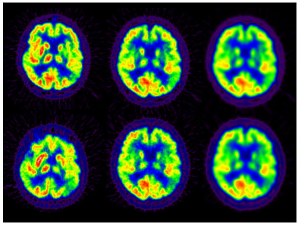
Methods: The Core has supervised the acquisition, quality control, and analysis of longitudinal [(18)F]fluorodeoxyglucose PET (FDG-PET) data in approximately half of the ADNI cohort. In an "add on" study, approximately 100 subjects also underwent scanning with [(11)C] Pittsburgh compound B PET for amyloid imaging. The Core developed quality control procedures and standardized image acquisition by developing an imaging protocol that has been widely adopted in academic and pharmaceutical industry studies. Data processing provides users with scans that have identical orientation and resolution characteristics despite acquisition on multiple scanner models. The Core labs have used many different approaches to characterize differences between subject groups (Alzheimer's disease, mild cognitive impairment, controls), to examine longitudinal change over time in glucose metabolism and amyloid deposition, and to assess the use of FDG-PET as a potential outcome measure in clinical trials.

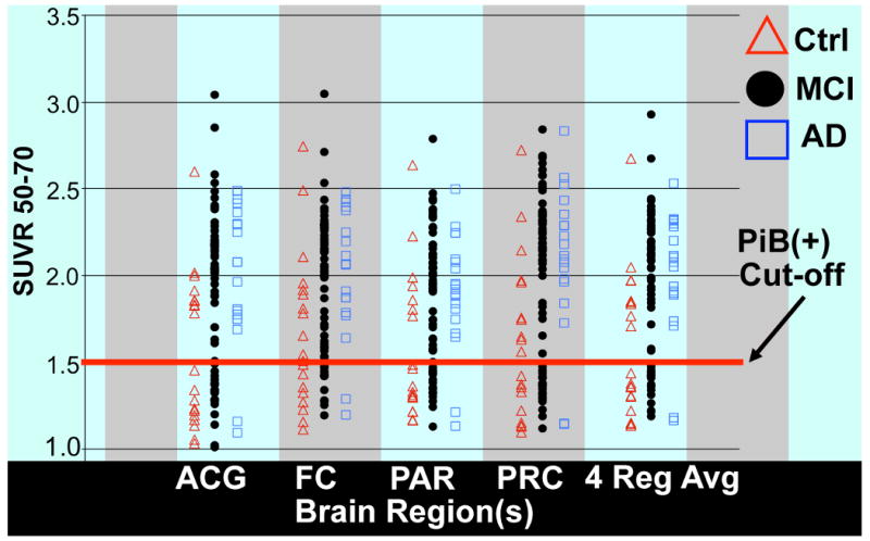
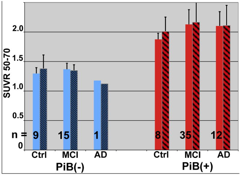
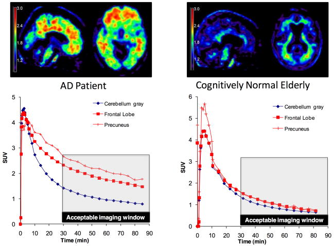
Results: ADNI data indicate that FDG-PET increases statistical power over traditional cognitive measures, might aid subject selection, and could substantially reduce the sample size in a clinical trial. Pittsburgh compound B PET data showed expected group differences, and identified subjects with significant annual increases in amyloid load across the subject groups. The next activities of the PET core in ADNI will entail developing standardized protocols for amyloid imaging using the [(18)F]-labeled amyloid imaging agent AV45, which can be delivered to virtually all ADNI sites.

Conclusions: ADNI has demonstrated the feasibility and utility of multicenter PET studies and is helping to clarify the role of biomarkers in the study of aging and dementia.


Conflict of interest statement

GE Healthcare holds a license agreement with the University of Pittsburgh based on the PIB technology described in this manuscript. Dr. Mathis is a co-inventor of PIB and, as such, has a financial interest in this license agreement. GE Healthcare had no role in the design or interpretation of results or preparation of this manuscript.

Figures

Figure 1
Figure 1
Examples of the ADNI processing stream. Top row: Baseline FDG scan, MCI subject. Bottom row: 36m FDG scan, same subject. Left: FDG-PET image in native format and orientation, averaged 6 × 5 min frames. Middle: Same as left but with standardized orientation, image grid, and voxel size. Right: Same as middle but smoothed to an isotropic resolution of 8 mm FWHM. Note longitudinal decrease seen in left parietal cortex (image right) in middle and right columns.
Figure 2
Figure 2
These regions of interest (ROIs) were generated as described in the text using clusters of significant voxels from the FDG-PET literature. These were averaged together into a composite ROI for the ROI-based analyses.
Figure 3
Figure 3
Results of SPM analyses showing significant 12 month declines in glucose metabolism in patients with AD (A) and MCI (B).
Figure 4
Figure 4
SSP Images in ADNI MCI subjects by diagnosis at 12 months after imaging.
Figure 5
Figure 5
Regional PIB-PET SUVR outcome measure values in four brain regions (ACG, anterior cingulate; FC, frontal cortex; PAR, parietal cortex; and PRC, precuneus) and a 4 cortical regions averaged value (4 Reg Avg) for all 103 PIB-PET baseline subjects in the ADNI PIB add-on studies. The cut-off value of 1.50 SUVR units dichotomizes all of the subjects into two groups: PIB(+) and PIB(-). Subjects are normal (red triangles), AD (blue boxes) and MCI (black circles).
Figure 6
Figure 6
Four Region Average SUVR values for 80 PIB-PET one year longitudinal follow-up subjects separated into control, MCI, and AD groups and each group further sub-divided into PIB(-) and PIB(+) sub-groups. n is the number of subjects in each sub-group and hatched boxes indicate the one year follow-up sub-group SUVR averages.
Figure 7
Figure 7
Top: 10 min PET data from AV-45 in an AD patient (left) and healthy control (right) showing uptake in cortex in AD and nonspecific retention in white matter in the control. Bottom: Kinetics that demonstrate rapid uptake and washout with retention in cortical regions in the AD patient, washout in the same regions in controls, and similar washout in white matter and cerebellum in both subjects.

Similar articles

Cited by

References

    1. Joshi A, Koeppe RA, Fessler JA. Reducing between scanner differences in multi-center PET studies. Neuroimage. 2009;46:154–159. - PMC - PubMed
    1. Minoshima S, Frey KA, Koeppe RA, Foster NL, Kuhl DE. A diagnostic approach in Alzheimer's disease using three-dimensional stereotactic surface projections of fluorine-18-FDG PET. J Nucl Med. 1995;36:1238–1248. - PubMed
    1. Landau SM, Harvey D, Madison CM, Koeppe RA, Reiman EM, Foster NL, et al. Associations between cognitive, functional, and FDG-PET measures of decline in AD and MCI. Neurobiol Aging. 2009 - PMC - PubMed
    1. Jagust WJ, Landau SM, Shaw LM, Trojanowski JQ, Koeppe RA, Reiman EM, et al. Relationships between biomarkers in aging and dementia. Neurology. 2009;73:1193–1199. - PMC - PubMed
    1. Langbaum JB, Chen K, Lee W, Reschke C, Bandy D, Fleisher AS, et al. Categorical and correlational analyses of baseline fluorodeoxyglucose positron emission tomography images from the Alzheimer's Disease Neuroimaging Initiative (ADNI) Neuroimage. 2009;45:1107–1116. - PMC - PubMed

Publication types