

Histological types of breast cancer: how special are they?
- PMID: 20452298
- PMCID: PMC5527938
- DOI: 10.1016/j.molonc.2010.04.004
Histological types of breast cancer: how special are they?
Abstract
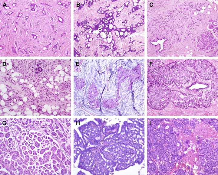
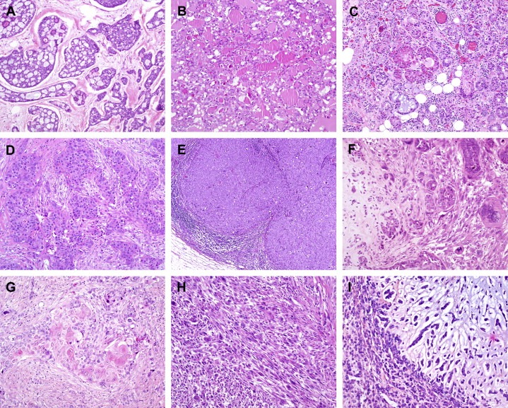
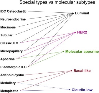
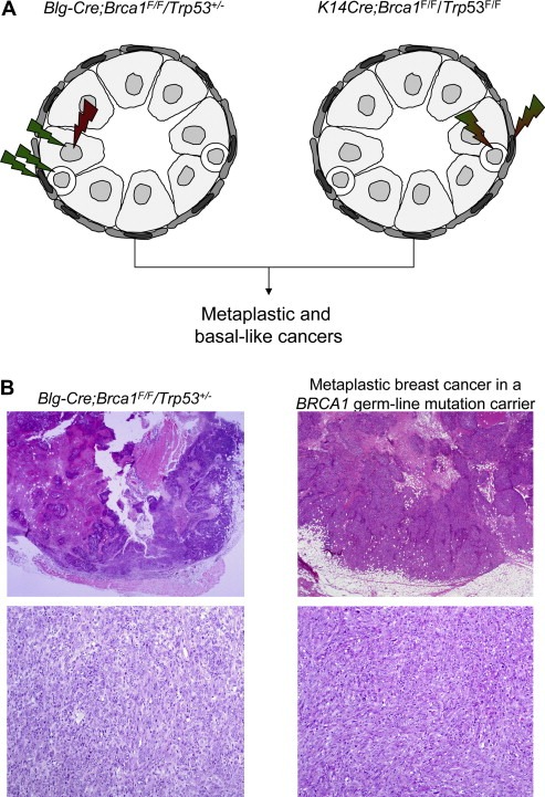
Breast cancer is a heterogeneous disease, comprising multiple entities associated with distinctive histological and biological features, clinical presentations and behaviours and responses to therapy. Microarray-based technologies have unravelled the molecular underpinning of several characteristics of breast cancer, including metastatic propensity and histological grade, and have led to the identification of prognostic and predictive gene expression signatures. Furthermore, a molecular taxonomy of breast cancer based on transcriptomic analysis has been proposed. However, microarray studies have primarily focused on invasive ductal carcinomas of no special type. Owing to the relative rarity of special types of breast cancer, information about the biology and clinical behaviour of breast cancers conveyed by histological type has not been taken into account. Histological special types of breast cancer account for up to 25% of all invasive breast cancers. Recent studies have provided direct evidence of the existence of genotypic-phenotypic correlations. For instance, secretory carcinomas of the breast consistently harbour the t(12;15) translocation that leads to the formation of the ETV6-NTRK3 fusion gene, adenoid cystic carcinomas consistently display the t(6;9) MYB-NFIB translocation and lobular carcinomas consistently show inactivation of the CDH1 gene through multiple molecular mechanisms. Furthermore, histopathological and molecular analysis of tumours from conditional mouse models has provided direct evidence for the causative role of specific genes in the genesis of specific histological special types of breast cancer. Here we review the associations between the molecular taxonomy of breast cancer and histological special types, discuss the possible origins of the heterogeneity of breast cancer and propose an approach for the identification of novel therapeutic targets based on the study of histological special types of breast cancer.
(c) 2010 Federation of European Biochemical Societies. Published by Elsevier B.V. All rights reserved.
Figures
References
-
- Abd El-Rehim, D.M. , Pinder, S.E. , Paish, C.E. , Bell, J. , Blamey, R.W. , Robertson, J.F. , Nicholson, R.I. , Ellis, I.O. , 2004. Expression of luminal and basal cytokeratins in human breast carcinoma. J. Pathol. 203, 661–671. - PubMed
-
- Abdel-Fatah, T.M. , Powe, D.G. , Hodi, Z. , Lee, A.H. , Reis-Filho, J.S. , Ellis, I.O. , 2007. High frequency of coexistence of columnar cell lesions, lobular neoplasia, and low grade ductal carcinoma in situ with invasive tubular carcinoma and invasive lobular carcinoma. Am. J. Surg. Pathol. 31, 417–426. - PubMed
-
- Abdel-Fatah, T.M. , Powe, D.G. , Hodi, Z. , Reis-Filho, J.S. , Lee, A.H. , Ellis, I.O. , 2008. Morphologic and molecular evolutionary pathways of low nuclear grade invasive breast cancers and their putative precursor lesions: further evidence to support the concept of low nuclear grade breast neoplasia family. Am. J. Surg. Pathol. 32, 513–523. - PubMed
-
- Adelaide, J. , Finetti, P. , Bekhouche, I. , Repellini, L. , Geneix, J. , Sircoulomb, F. , Charafe-Jauffret, E. , Cervera, N. , Desplans, J. , Parzy, D. , Schoenmakers, E. , Viens, P. , Jacquemier, J. , Birnbaum, D. , Bertucci, F. , Chaffanet, M. , 2007. Integrated profiling of basal and luminal breast cancers. Cancer Res. 67, 11565–11575. - PubMed
Publication types
MeSH terms
Substances
Grants and funding
LinkOut - more resources
Full Text Sources
Medical
Miscellaneous
