

Addition of glutamate to serum-free culture promotes recovery of electrical activity in adult hippocampal neurons in vitro
- PMID: 20452373
- PMCID: PMC2925239
- DOI: 10.1016/j.jneumeth.2010.04.030
Addition of glutamate to serum-free culture promotes recovery of electrical activity in adult hippocampal neurons in vitro
Abstract
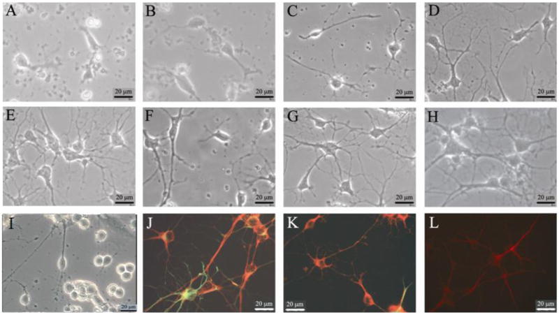
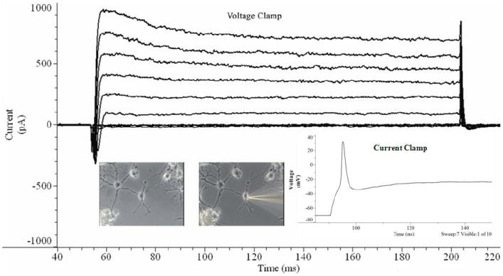
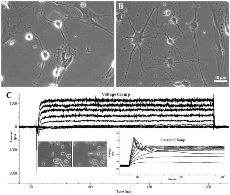
A long-term cell culture system utilizing normal adult hippocampal neurons would represent an important tool that could be useful in research on the mature brain, neurological disorders and age-related neurological diseases. Historically, in vitro neuronal systems are derived from embryonic rather than mature brain tissue, a practice predicated upon difficulties in supporting regeneration, functional recovery and long-term survival of adult neurons in vitro. A few studies have shown that neurons derived from the hippocampal tissue of adult rats can survive and regenerate in vitro under serum-free conditions. However, while the adult neurons regenerated morphologically under these conditions, both the electrical activity characteristic of in vivo neurons as well as long-term neuronal survival was not consistently recovered in vitro. In this study, we report on the development of a defined culture system with the ability to support functional recovery and long-term survival of adult rat hippocampal neurons. In this system, the cell-adhesive substrate, N-1 [3-(trimethoxysilyl) propyl]-diethylenetriamine, supported neuronal attachment, regeneration, and long-term survival of adult neurons for more than 80 days in vitro. Additionally, the excitatory neurotransmitter glutamate, applied at 25muM for 1-7 days after morphological neuronal regeneration in vitro, enabled full recovery of neuronal electrical activity. This low concentration of glutamate promoted the recovery of neuronal electrical activity but with minimal excitotoxicity. These improvements allowed electrically active adult neurons to survive in vitro for several months, providing a stable test-bed for the long-term study of regeneration in adult-derived neuronal systems, especially for traumatic brain injury (TBI).
Copyright 2010 Elsevier B.V. All rights reserved.
Figures
References
-
- Auditore JV, Wade L, Olson EJ. Occurrence of N-acetyl-L-glutamic acid in the human brain. Journal of Neurochemistry. 1966;13(11):1149–1155. - PubMed
-
- Balazs R. Trophic effect of glutamate. Curr Top Med Chem. 2006;6(10):961–968. - PubMed
-
- Barone FC, White RF, Spera PA, Ellison J, Currie RW, Wang X, Feuerstein GZ, Rothwell NJ. Ischemic Preconditioning and Brain Tolerance: Temporal Histological and Functional Outcomes, Protein Synthesis Requirement, and Interleukin-1 Receptor Antagonist and Early Gene Expression. Stroke. 1998;29(9):1937–1951. - PubMed
-
- Bousse L. Whole cell biosensors. Sensors and Actuators B-Chemical. 1996;34(1-3):270–275.
-
- Brewer GJ. Isolation and culture of adult rat hippocampal neurons. J Neurosci Methods. 1997;71(2):143–55. - PubMed
Publication types
MeSH terms
Substances
Grants and funding
LinkOut - more resources
Full Text Sources
Other Literature Sources
