

Changes in the frontotemporal cortex and cognitive correlates in first-episode psychosis
- PMID: 20452574
- PMCID: PMC3025327
- DOI: 10.1016/j.biopsych.2010.03.019
Changes in the frontotemporal cortex and cognitive correlates in first-episode psychosis
Abstract
Background: Loss of cortical volume in frontotemporal regions has been reported in patients with schizophrenia and their relatives. Cortical area and thickness are determined by different genetic processes, and measuring these parameters separately may clarify disturbances in corticogenesis relevant to schizophrenia. Our study also explored clinical and cognitive correlates of these parameters.
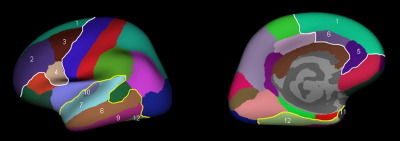
Methods: Thirty-seven patients with first-episode psychosis (34 schizophrenia, 3 schizoaffective disorder) and 38 healthy control subjects matched for age and sex took part in the study. Imaging was performed on an magnetic resonance imaging 1.5-T scanner. Area and thickness of the frontotemporal cortex were measured using a surface-based morphometry method (Freesurfer). All subjects underwent neuropsychologic testing that included measures of premorbid and current IQ, working and verbal memory, and executive function.
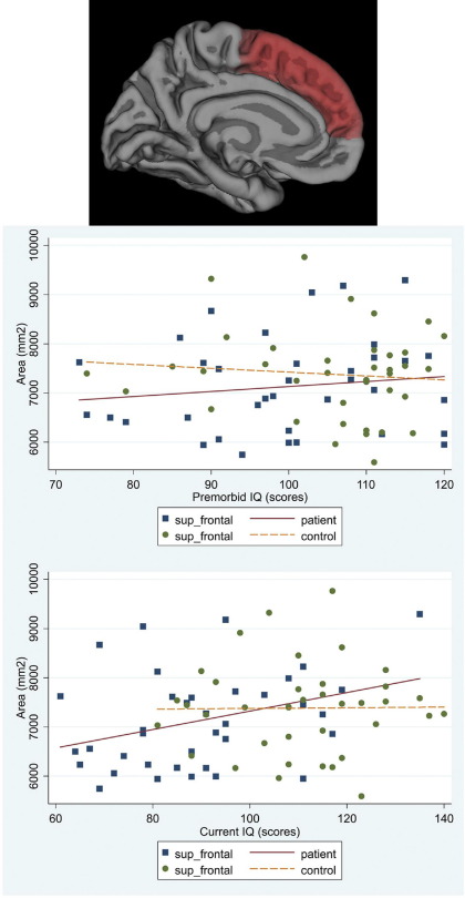
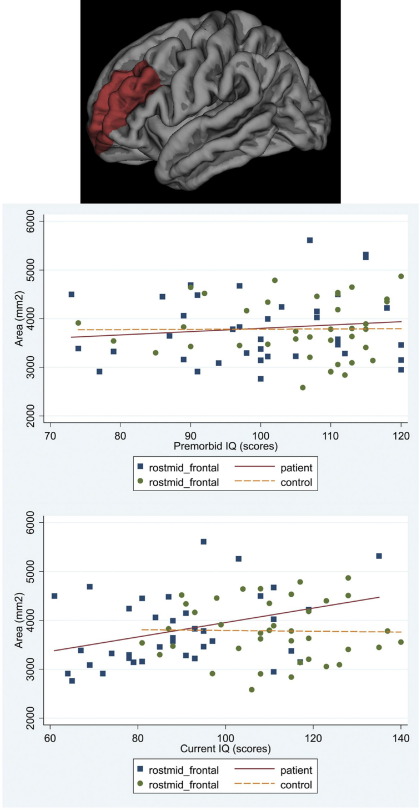
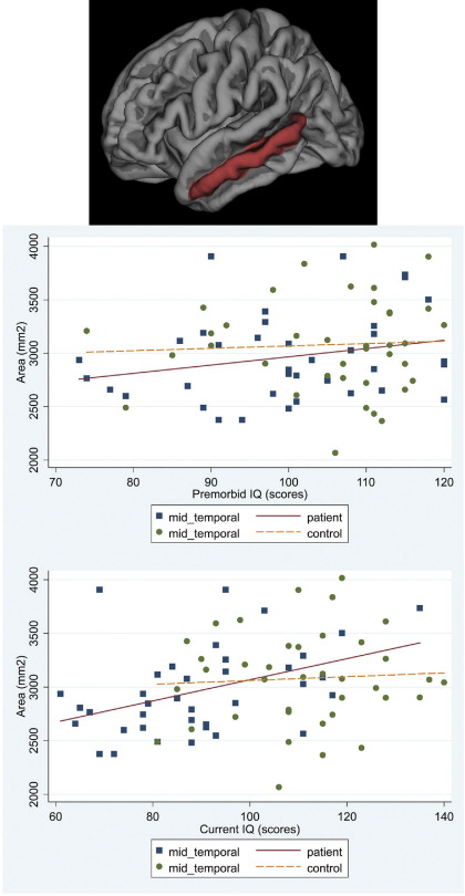
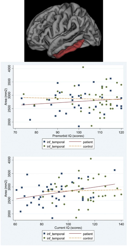
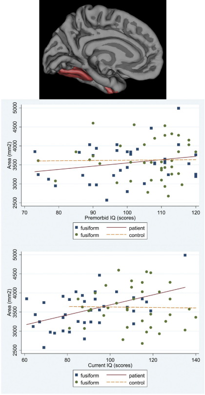
Results: Reductions in cortical area, more marked in the temporal cortex, were present in patients. Overall frontotemporal cortical thickness did not differ between groups, although regional thinning of the right superior temporal region was observed in patients. There was a significant association of both premorbid IQ and IQ at disease onset with area, but not thickness, of the frontotemporal cortex, and working memory span was associated with area of the frontal cortex. These associations remained significant when only patients with schizophrenia were considered.
Conclusions: Our results suggest an early disruption of corticogenesis in schizophrenia, although the effect of subsequent environmental factors cannot be excluded. In addition, cortical abnormalities are subject to regional variations and differ from those present in neurodegenerative diseases.
Copyright 2010 Society of Biological Psychiatry. Published by Elsevier Inc. All rights reserved.
Figures
Similar articles
-
IQ and the fronto-temporal cortex in bipolar disorder.J Int Neuropsychol Soc. 2012 Mar;18(2):370-4. doi: 10.1017/S1355617711001706. Epub 2012 Jan 23. J Int Neuropsychol Soc. 2012. PMID: 22264359
-
Cortical abnormalities and their cognitive correlates in patients with temporal lobe epilepsy and interictal psychosis.Epilepsia. 2012 Jun;53(6):1077-87. doi: 10.1111/j.1528-1167.2012.03504.x. Epub 2012 May 11. Epilepsia. 2012. PMID: 22578165
-
A longitudinal study of cortical changes and their cognitive correlates in patients followed up after first-episode psychosis.Psychol Med. 2015 Jan;45(1):205-16. doi: 10.1017/S0033291714001433. Epub 2014 Jul 3. Psychol Med. 2015. PMID: 24990283
-
Cortical thickness reduction in individuals at ultra-high-risk for psychosis.Schizophr Bull. 2011 Jul;37(4):839-49. doi: 10.1093/schbul/sbp151. Epub 2009 Dec 21. Schizophr Bull. 2011. PMID: 20026559 Free PMC article.
-
Subjective rating of working memory is associated with frontal lobe volume in schizophrenia.Schizophr Res. 2010 Jul;120(1-3):71-5. doi: 10.1016/j.schres.2010.02.1067. Epub 2010 Mar 20. Schizophr Res. 2010. PMID: 20303715 Free PMC article.
Cited by
-
Gray matter changes and cognitive predictors of 2-year follow-up abnormalities in early-onset first-episode psychosis.Eur Child Adolesc Psychiatry. 2018 Jan;27(1):113-126. doi: 10.1007/s00787-017-1013-z. Epub 2017 Jul 13. Eur Child Adolesc Psychiatry. 2018. PMID: 28707138
-
A window into the brain: an in vivo study of the retina in schizophrenia using optical coherence tomography.Psychiatry Res. 2012 Jul 30;203(1):89-94. doi: 10.1016/j.pscychresns.2011.08.011. Epub 2012 Aug 20. Psychiatry Res. 2012. PMID: 22917503 Free PMC article.
-
Effects of the neurogranin variant rs12807809 on thalamocortical morphology in schizophrenia.PLoS One. 2013 Dec 30;8(12):e85603. doi: 10.1371/journal.pone.0085603. eCollection 2013. PLoS One. 2013. PMID: 24386483 Free PMC article. Clinical Trial.
-
Distinct abnormalities of the primate prefrontal cortex caused by ionizing radiation in early or midgestation.J Comp Neurol. 2013 Apr 1;521(5):1040-53. doi: 10.1002/cne.23217. J Comp Neurol. 2013. PMID: 22911497 Free PMC article.
-
Brain functional connectivity associated with cognitive deficits in younger patients at first episode of schizophrenia.Schizophr Res Cogn. 2025 Mar 31;41:100359. doi: 10.1016/j.scog.2025.100359. eCollection 2025 Sep. Schizophr Res Cogn. 2025. PMID: 40567506 Free PMC article.
References
-
- Friston K.J., Frith C.D. Schizophrenia: A disconnection syndrome. Clin Neurosci. 1995;3:89–97. - PubMed
-
- Harrison P.J., Weinberger D.R. Schizophrenia genes, gene expression, and neuropathology: On the matter of their convergence. Mol Psychiatry. 2005;10:40–68. - PubMed
-
- Honea R., Crow T.J., Passingham D., Mackay C.E. Regional deficits in brain volume in schizophrenia: A meta-analysis of voxel-based morphometry studies. Am J Psychiatry. 2005;162:2233–2245. - PubMed
Publication types
MeSH terms
Grants and funding
LinkOut - more resources
Full Text Sources
Medical
