. 2010 Dec;38(6):691-8.
doi: 10.1016/j.ejcts.2010.03.049. Epub 2010 May 10.

Regenerative healing following foetal myocardial infarction

Affiliations

Regenerative healing following foetal myocardial infarction

Benjamin J Herdrich et al. Eur J Cardiothorac Surg. 2010 Dec.

Abstract

Objectives: The adult response to myocardial infarction results in inflammation, scar formation, left ventricular dilatation, and loss of regional and global function. Regenerative scarless healing has been demonstrated in foetal dermis and tendon and is associated with diminished inflammation. We hypothesised that following foetal myocardial infarction, there would be minimal inflammation, regenerative healing, and preservation of function.

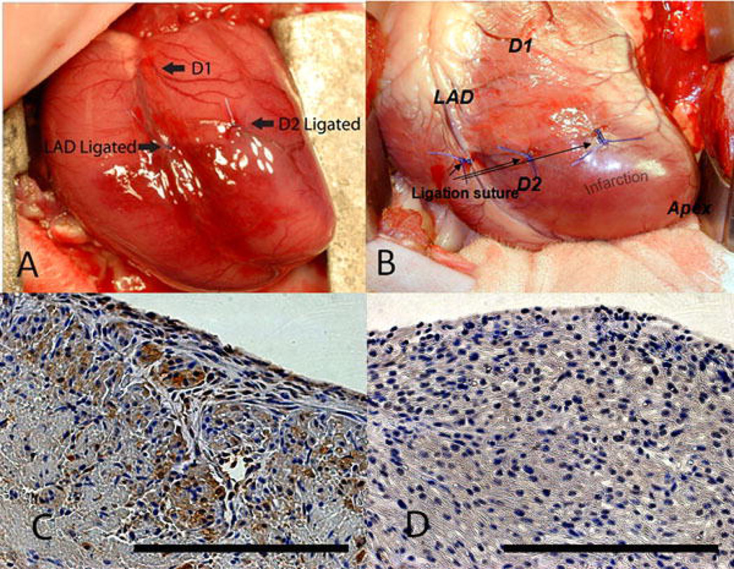
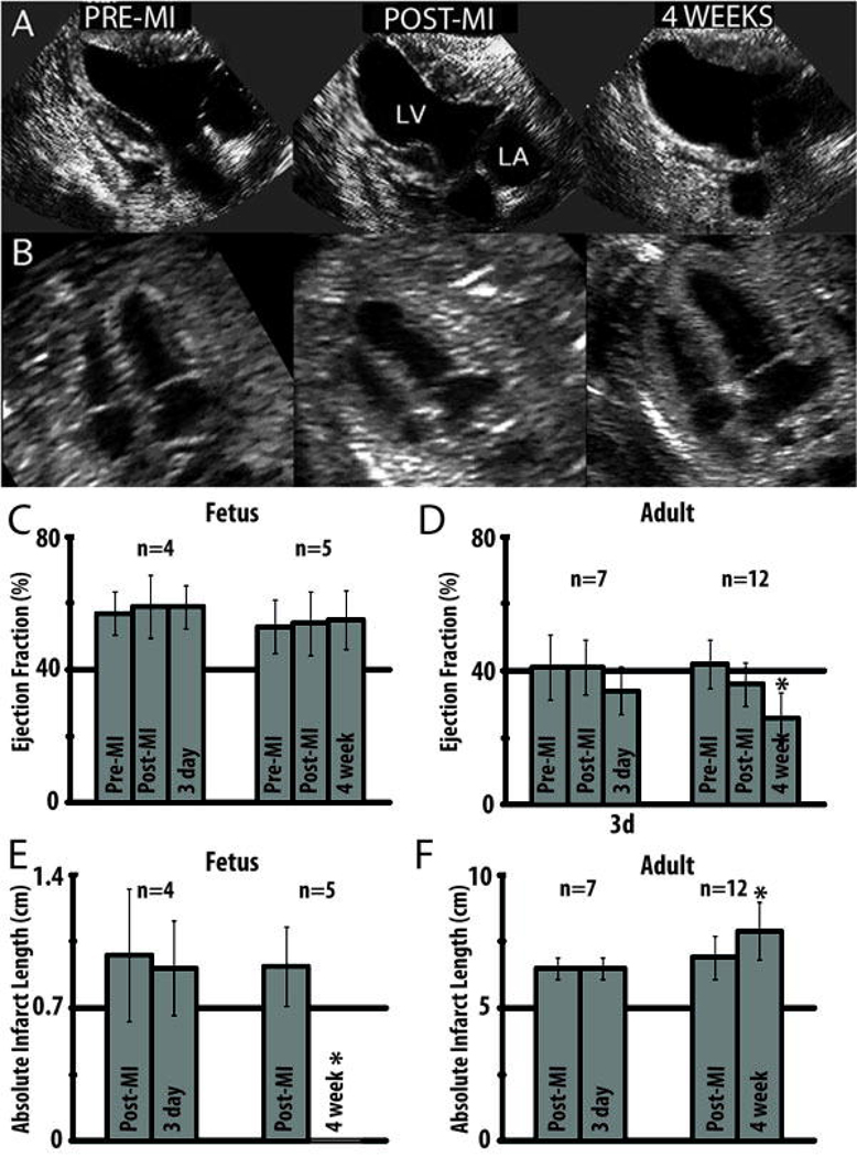
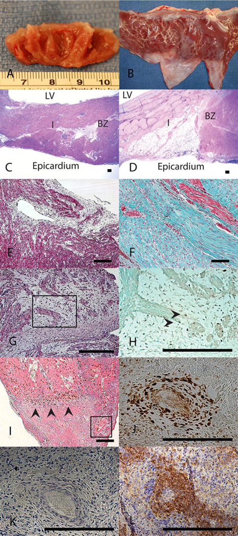
Methods: Anteroapical myocardial infarction encompassing 20% of the left ventricle was created in adult or early gestation foetal sheep. Myocardial function was serially assessed using quantitative echocardiography. Infarct architecture was examined histologically for evidence of scar formation. Cellular inflammation, cellular proliferation, and apoptosis were assessed using immunohistochemistry.

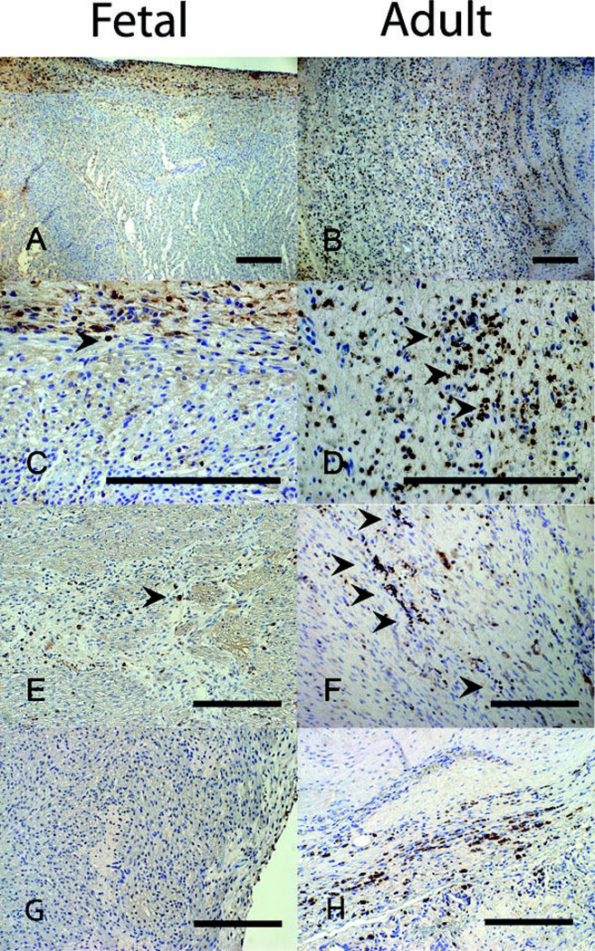
Results: In the adult sheep 4 weeks following myocardial infarction, there was a significant decline in ejection fraction (EF) (41±7.4% to 26±7.4%, p<0.05), and the akinetic myocardial segment increased in size (6.9±0.8 cm to 7.9±1.1 cm, p<0.05). By contrast, there was no decline in the foetal EF (53±8.1% to 55±8.8%) and no akinetic foetal myocardial segment 4 weeks post-infarction. The foetal infarcts lacked an inflammatory cell infiltrate and healed with minimal fibrosis, compared with the adults. Foetal infarcts also demonstrated 5-bromo-2'-deoxyuridine (BrdU)+ proliferating cells, including cardiomyocytes, within the infarct.

Conclusions: These data demonstrate that the foetal response to myocardial infarction is dramatically different from the adult and is characterised by minimal inflammation, lack of fibrosis, myocardial proliferation and restoration of cardiac function. Diminished inflammation is associated with foetal regenerative cardiac healing following injury. Understanding the mechanisms involved in foetal myocardial regeneration may lead to applications to alter the adult response following myocardial infarction.


Figures

Figure 1
Figure 1
Ligation of the distal LAD and D2 leads to a reproducible infarct in fetal and adult hearts. An anteroapical MI is created by ligating the LAD and D2 at a point 40% of the distance between the apex and the base in (a) fetal and (b) adult sheep. Activated caspase-3 staining of fetal hearts 3 days following infarction demonstrates (c) apoptosis in the area of infarction (400×) and (d) lack of apoptosis away from the area of infarction (400×). Scale bars equal 200µm.
Figure 2
Figure 2
Echocardioghraphic assessment of fetal and adult hearts following myocardial infarction demonstrates functional decline in the adult and restoration of function in the fetus. (a) Serial end-systolic echocardiographic views demonstrate dilation of the LV, 4 weeks following infarction with a large anteroapical infarct in the adult. (b) In the fetus, there is no evidence of LV dilation or infarcted myocardium at 4 weeks. (c) EF measured by quantitative echocardiography is unchanged in the fetus at 3 days (p=0.37) and 4 weeks (p=0.31) following infarction. (d) In the adult, the EF has significantly declined by 4 weeks following myocardial infarction (*p<0.05 vs. adult pre-MI and post-MI). (e) Absolute infarct length defined as the length of akinetic myocardium measured by echocardiography is unchanged at 3 days following infarction (p=0.72) but decreases to zero in the fetus at 4 weeks following infarction (*p<0.05 vs fetal post-MI). (f) In the adult, the absolute infarct length is also unchanged at 3 days following infarction (p=1.00) but increases over a period of 4 weeks following infarction (*p<0.05 vs adult post-MI).
Figure 3
Figure 3
Fetal cardiac ventricular remodeling following MI proceeds with regeneration of myocardium and without scar formation. 4 weeks after myocardial infarction (a) fetal hearts show no gross evidence of fibrosis while (b) adult hearts show apical fibrosis and ventricular wall thinning. H&E staining at 4 weeks demonstrates (c) no evidence of myocyte loss or ventricular wall thinning in the fetal heart (I) infarct or (BZ) borderzone and (d) significant myocyte loss and ventricular wall thinning in the adult infarct (I) (20×). Masson’s trichrome staining at 4 weeks following MI confirms that there is (e) minimal fibrosis in the fetal infarct (100×) and (f) an exuberant fibrotic response in the adult infarct (100×). (g) 4 weeks following myocardial infarction, H&E staining of the fetal infarct shows disordered clusters of cells with minimal surrounding fibrosis suggesting myocardial regeneration (200×). (h) BrdU immunostaining with Lichgrün counterstain on a serial section demonstrates that within these clusters, there are cycling, BrdU positive, cardiomyocytes marked by the arrowheads confirming myocardial proliferation within the infarct (400×). (i) BrdU immunostaining with eosin counterstain on a fetal heart 3 days following MI demonstrates a polarity for BrdU positive cells which accumulate around the area of infarction (marked with arrowheads, 50× optical, 2× digital) and (j) surrounding blood vessels within the infarct (serial section with Lichgrün counterstain, 400×). (k) CD45 immunostaining on a serial section of this blood vessel within the fetal infarct demonstrates that these BrdU positive cells are not positive for CD45, indicating they are not of hematopoetic lineage. (l) Positive control of CD45 immunohistochemistry on a fetal sheep spleen. Scale bars equal 200µm.
Figure 4
Figure 4
CD45 immunmohistochemistry following myocardial infarction demonstrates markedly less cellular inflammatory response in fetal verses adult hearts. 7 days following infarction the fetal heart (a-100×, c-400×) shows minimal numbers of inflammatory cells while the adult heart (b-100×, d-400×) shows a large inflammatory infiltrate. At 4 weeks following infarction, the number of inflammatory cells in both the (e) fetal and (f) adult hearts has decreased, but the adult heart has persistent scattered areas of inflammation not seen in the fetus (200×). (g) Caspase-3 staining in the fetal heart 4 weeks after infarction fails to show any apoptosis (200×). (h) Caspase-3 staining in the adult infarct 4 weeks after infarction demonstrates continuing apoptosis (200×). Scale bars equal 200µm.

Similar articles

Cited by

References

    1. Yang Z, Day YJ, Toufektsian MC, Xu Y, Ramos SI, Marshall MA, French BA, Linden J. Myocardial infarct-sparing effect of adenosine A2A Receptor Activation is due to its action on CD4+ T lymphocytes. Circulation. 2006;114:2056–2064. - PubMed
    1. St. John Sutton M, Sharpe N. Left ventricular remodeling after infarction: pathophysiology and therapy. Circulation. 2000;101:2981–2988. - PubMed
    1. Cleutjens JPM, Kandala JC, Guarda E, Guntaka RV, Weber KT. Regulation of collagen degradation in the rat myocardium after infarction. J Mol Cell Cardiol. 1995;27:1281–1292. - PubMed
    1. Takahashi T, Hiasa Y, Ohara Y, Miyazaki S, Ogura R, Miyajima H, Yuba K, Suzuki N, Hosokawa S, Kishi K, Ohtani R. Relation between neutrophil counts on admission, microvascular injury, and left ventricular functional recovery in patients with an anterior wall first acute myocardial infarction treated with primary coronary angioplasty. Am J Cardiol. 2007;100:35–40. - PubMed
    1. Åströ-Olsson K, Hedström E, Hultén LM, Wiklund O, Arheden H, Öhlin AK, Gottsäter A, Öhlin H. Dissociation of the inflammatory reaction following PCI for acute myocardial infarction. J Invasive Cardiol. 2007;19:452–456. - PubMed

Publication types