

Case Reports
doi: 10.1186/2047-783x-15-2-84.
Giant plexiform neurofibroma with hemorrhage in cranio-maxillofacial region as depicted on CT and MRI
Affiliations
- PMID: 20452890
- PMCID: PMC3352051
- DOI: 10.1186/2047-783x-15-2-84
Item in Clipboard
Case Reports
Giant plexiform neurofibroma with hemorrhage in cranio-maxillofacial region as depicted on CT and MRI
Eur J Med Res.
.
Abstract
Plexiform neurofibroma (PN) is a rare benign tumor and a special subtype of neurofibromatosis type 1 (NF1). Though the incidence is low, giant PN of the craniomaxillofacial region could result in severe hemifacial hypertrophy which is known as a typical manifestation of NF1 in young children. Here, we retrospectively reported a giant plexiform neurofibroma with hemorrhage in the cranio-maxillofacial region detected by CT and MRI. In addition, a brief review of the relevant literature is presented.
Figures
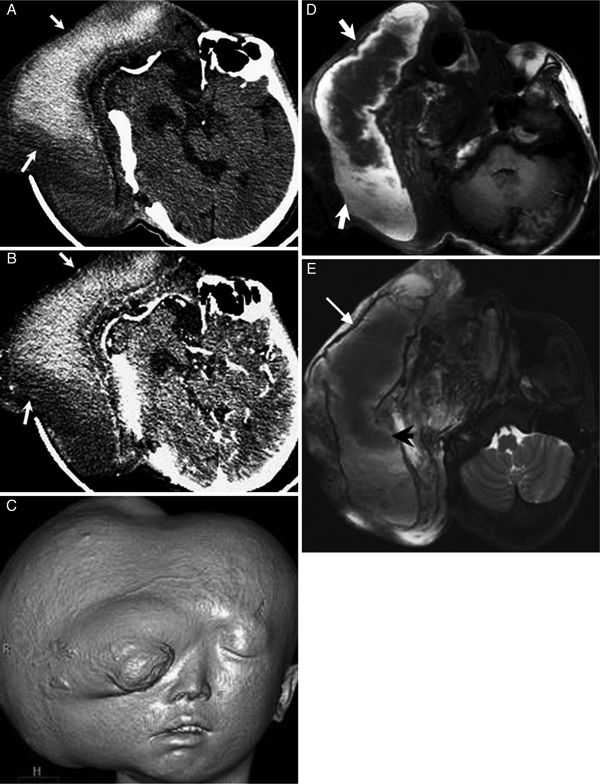
A 21-year-old man with plexiforma neurofibroma in the right cranio-maxillofacial region. (A) Non-enhanced CT demonstrates a giant cystic-solid mass in the right craniomaxillofacial region, and the cyst of the mass appearing as hyper- and iso- attenuation (white arrows). (B) Contrast-enhanced CT depicts the mass appearing as no enhancement (arrows). (C) SSD reconstruction CT shows the cosmetic impairment of right face. (D) On T1-weighted MR image, the cyst of the mass shows heterogeneously intermediate to high signal intensity (arrows). (E) The cyst of the mass shows light low to intermediate signal intensity on T2 weighted MR image (arrowhead) with a low-signal ring around the hematoma (thin arrows).
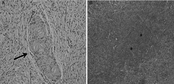
Photomicrography of the case mention in figure 1. (A) Histology specimen demonstrates that the tumor is consisted of spindle-like schwann cells (arrow) (HE 200 ×), and (B) the area of hemorrhage in the mass (*, HE 100 ×).
References
-
- Overdiek A, Feifel H, Schaper J, Mayatepek E, Rosenbaum T. Diagnostic delay of NF1 in hemifacial hypertrophy due to plexiform neurofibromas. No To Hattasu (Brain and Development) 2006;28(5):275–280. - PubMed
-
- Hersh JH. Health supervision for children with neurofibromatosis. Pediatrics. 2008;21:633–642. - PubMed
-
- Malagari K, Drakopoulos S, Brountzos E, Sissopulos A, Efthimidadou A, Hadjiyiannakis E. et al.Plexiform neurofibroma of the liver: findings on MR imaging, angiography, and CT portography. AJR. 2001;176:493–495. - PubMed
Publication types
MeSH terms
LinkOut - more resources
Full Text Sources
Medical
Research Materials
Miscellaneous
