

Optical coherence tomography for bladder cancer -- ready as a surrogate for optical biopsy? Results of a prospective mono-centre study
- PMID: 20452899
- PMCID: PMC3352220
- DOI: 10.1186/2047-783x-15-3-131
Optical coherence tomography for bladder cancer -- ready as a surrogate for optical biopsy? Results of a prospective mono-centre study
Abstract
Introduction: New modalities like Optical Coherence Tomography (OCT) allow non-invasive examination of the internal structure of biological tissue in vivo. The potential benefits and limitations of this new technology for the detection and evaluation of bladder cancer were examined in this study.
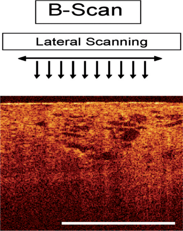
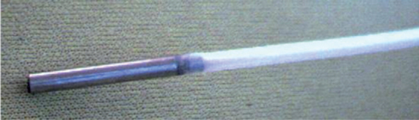
Materials and methods: Between January 2007 and January 2008, 52 patients who underwent transurethral bladder biopsy or TUR-BT for surveillance or due to initial suspicion of urothelial carcinoma of the bladder were enrolled in this study. In total, 166 lesions were suspicious for malignancy according to standard white light cystoscopy. All suspicious lesions were scanned and interpreted during perioperative cystoscopy using OCT. Cold cup biopsies and/or TUR-B was performed for all these lesions. For this study we used an OCT-device (Niris, Imalux, Cleveland, US), that utilizes near-infrared light guided through a flexible fibre-based applicator, which is placed into the bladder via the working channel of the cystoscope. The technology provides high spatial resolution on the order of about 10-20 microm, and a visualization of tissue to a depth of about 2 mm across a lateral span of about 2 mm in width. The device used received market clearance from the FDA and CE approval in Germany. The diagnostic and surgical procedure was videotaped and analyzed afterwards for definitive matching of scanned and biopsied lesion. The primary aim of this study was to determine the level of correlation between OCT interpretation and final histological result.
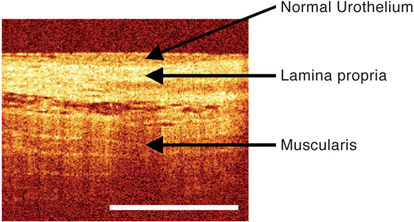
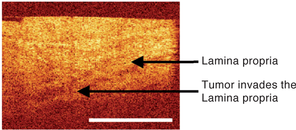
Results: Of 166 scanned OCT images, 102 lesions (61.4%) matched to the same site where the biopsy/TUR-BT was taken according to videoanalysis. Only these video-verified lesions were used for further analysis. Of all analyzed lesions 88 were benign (inflammation, edema, hyperplasia etc.) and 14 were malignant (CIS, Ta, T1, T2) as shown by final histo?pathology. - All 14 malignant lesions were detected correctly by OCT. Furthermore all invasive tumors were staged correctly by OCT regarding tumor growth beyond the lamina propria. There were no false negative lesions detected by OCT. Sensitivity of OCT for detecting the presence of a malignant lesion was 100% and sensitivity for detection of tumor growth beyond the lamina propria was 100% as well. Specificity of OCT for presence of malignancy was 65%, due to the fact that a number of lesions were interpreted as false positive by OCT.
Conclusion: As a minimally invasive technique, OCT proved to have extremely high sensitivity for detection of malignant lesions as well as estimation of whether a tumor has invaded beyond the lamina propria. However, specificity of OCT within the bladder was impaired (65%), possibly due to a learning curve and/or the relatively low spatial resolution and visualization depth of the OCT technology. Further studies and technical development are needed to establish an adequate surrogate for optical biopsy.
Figures
References
Publication types
MeSH terms
LinkOut - more resources
Full Text Sources
Medical
Research Materials
