Review
. 2010 Apr;2(4):a001842.
doi: 10.1101/cshperspect.a001842. Epub 2010 Mar 10.

Guidance molecules in synapse formation and plasticity

Affiliations
Review

Guidance molecules in synapse formation and plasticity

Kang Shen et al. Cold Spring Harb Perspect Biol. 2010 Apr.

Abstract

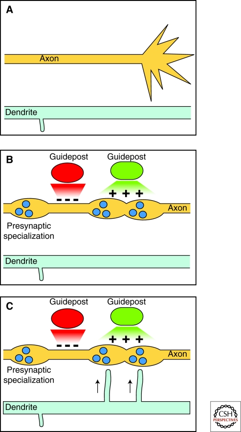
A major goal of modern neuroscience research is to understand the cellular and molecular processes that control the formation, function, and remodeling of chemical synapses. In this article, we discuss the numerous studies that implicate molecules initially discovered for their functions in axon guidance as critical regulators of synapse formation and plasticity. Insights from these studies have helped elucidate basic principles of synaptogenesis, dendritic spine formation, and structural and functional synapse plasticity. In addition, they have revealed interesting dual roles for proteins and cellular mechanisms involved in both axon guidance and synaptogenesis. Much like the dual involvement of morphogens in early cell fate induction and axon guidance, many guidance-related molecules continue to play active roles in controlling the location, number, shape, and strength of neuronal synapses during development and throughout the lifetime of the organism. This article summarizes key findings that link axon guidance molecules to specific aspects of synapse formation and plasticity and discusses the emerging relationship between the molecular and cellular mechanisms that control both axon guidance and synaptogenesis.


Figures

Figure 1.
Figure 1.
Basic steps of axo-dendritic synaptogenesis. Diagrams illustrate key steps involved in forming generic central synapses. (A) A guiding axon and nearby dendrite interact via cell–cell contacts mediated by the growth cone, collateral axon branches, or dendritic filopodial extensions. (B1) In some scenarios, pre-established presynaptic specializations mark the location of future synapses, whereas inappropriate axonal regions do not allow for synapse formation. (B2) In another scenario, random physical interactions between axon and dendrite (red boxes) form transient cell–cell adhesions. If the contacts are stable, then presynaptic and postsynaptic proteins, vesicles, and ion channels are recruited to the contact site. (C1 and C2) Stable contacts are matured into functional and morphological chemical synapses (spine or shaft synapses).
Figure 2.
Figure 2.
Prepatterning of synaptic specializations. Diagrams show the time-course of one form of synaptogenesis for en passant synapses. (A) An undifferentiated axon and dendrite before synapse formation. (B) Extrinsic cues promote (green) and inhibit (red) local presynapse formation, thereby patterning the subcellular localization of presynaptic terminals. (C) Postsynaptic dendritic filopodia approach presynaptic specializations to form synapses.
Figure 3.
Figure 3.
Guidance molecules involved in synapse formation and axon branching. (A) Model for EphB2 receptor functions and signaling in synaptogenesis. (B) Model for BDNF/TrkB receptor functions and signaling in synaptogenesis. (C) Role of ephrinBs in shaft and spine synapse formation.
Figure 4.
Figure 4.
Model for ephrinB, EphB, and BDNF/TrkB signaling in structural and functional synapse plasticity in the hippocampus.

Similar articles

Cited by

References

    1. Abe K, Chisaka O, Van Roy F, Takeichi M 2004. Stability of dendritic spines and synaptic contacts is controlled by α N-catenin. Nat Neurosci 7:357–363 - PubMed
    1. Alonso M, Medina JH, Pozzo-Miller L 2004. ERK1/2 activation is necessary for BDNF to increase dendritic spine density in hippocampal CA1 pyramidal neurons. Learn Mem 11:172–178 - PMC - PubMed
    1. Aoto J, Ting P, Maghsoodi B, Xu N, Henkemeyer M, Chen L 2007. Postsynaptic ephrinB3 promotes shaft glutamatergic synapse formation. J Neurosci 27:7508–7519 - PMC - PubMed
    1. Armstrong JN, Saganich MJ, Xu NJ, Henkemeyer M, Heinemann SF, Contractor A 2006. B-ephrin reverse signaling is required for NMDA-independent long-term potentiation of mossy fibers in the hippocampus. J Neurosci 26:3474–3481 - PMC - PubMed
    1. Augsburger A, Schuchardt A, Hoskins S, Dodd J, Butler S 1999. BMPs as mediators of roof plate repulsion of commissural neurons. Neuron 24:127–141 - PubMed

LinkOut - more resources