Review
. 2010 Apr;2(4):a001925.
doi: 10.1101/cshperspect.a001925.

Initiating and growing an axon

Affiliations
Review

Initiating and growing an axon

F Polleux et al. Cold Spring Harb Perspect Biol. 2010 Apr.

Abstract

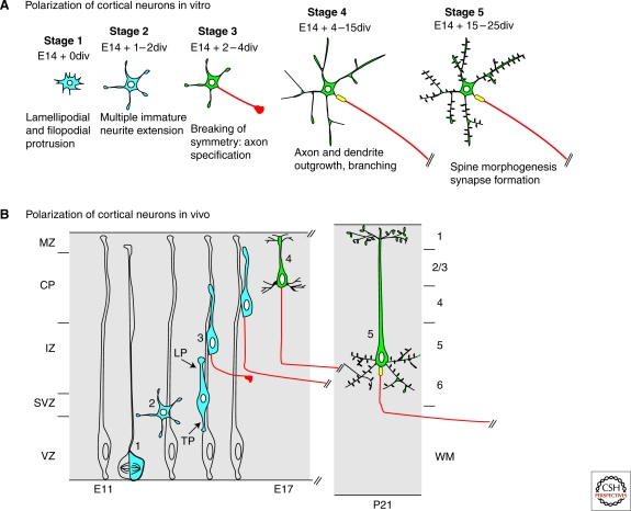
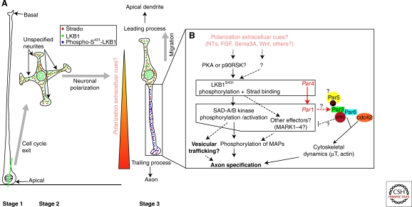
The ability of neurons to form a single axon and multiple dendrites underlies the directional flow of information transfer in the central nervous system. Dendrites and axons are molecularly and functionally distinct domains. Dendrites integrate synaptic inputs, triggering the generation of action potentials at the level of the soma. Action potentials then propagate along the axon, which makes presynaptic contacts onto target cells. This article reviews what is known about the cellular and molecular mechanisms underlying the ability of neurons to initiate and extend a single axon during development. Remarkably, neurons can polarize to form a single axon, multiple dendrites, and later establish functional synaptic contacts in reductionist in vitro conditions. This approach became, and remains, the dominant model to study axon initiation and growth and has yielded the identification of many molecules that regulate axon formation in vitro (Dotti et al. 1988). At present, only a few of the genes identified using in vitro approaches have been shown to be required for axon initiation and outgrowth in vivo. In vitro, axon initiation and elongation are largely intrinsic properties of neurons that are established in the absence of relevant extracellular cues. However, the importance of extracellular cues to axon initiation and outgrowth in vivo is emerging as a major theme in neural development (Barnes and Polleux 2009). In this article, we focus our attention on the extracellular cues and signaling pathways required in vivo for axon initiation and axon extension.


Figures

Figure 1.
Figure 1.
Parallels between neuronal polarization in vitro and in vivo. Comparison of the sequence of events leading to the polarization of cortical pyramidal neurons in vivo and in vitro. (A) In dissociated cultures, postmitotic cortical neurons display specific transitions as classically described for hippocampal neurons by Dotti and Banker (1988). At stage 1, immature postmitotic neurons display intense lamellipodial and filopodial protrusive activity, which leads to the emergence of multiple immature neurites, stage 2. Stage 3 represents a critical step when neuronal symmetry breaks and a single neurite grows rapidly to become the axon (purple), whereas other neurites acquire dendritic identity. Stage 4 is characterized by rapid axon and dendritic outgrowth. Finally, stage 5 neurons are terminally differentiated pyramidal neurons harboring dendritic spines and the AIS. (B) The axon–dendrite polarity of pyramidal neurons is derived from the polarized emergence of the trailing (TP) and leading processes (LP), respectively. In vivo, pyramidal neurons acquire other key features of their terminal polarity, such as the axon initiation segment (AIS; yellow cartridge) and dendritic spines (gray protrusions) during the first postnatal weeks of development.
Figure 2.
Figure 2.
Molecular mechanisms underlying cortical neuron polarization in vivo. (A) On asymmetric cell division of radial glial progenitors (Stage 1), early unpolarized postmitotic neurons show a transient phase of nondirected neurite outgrowth in the subventricular zone (Stage 2) before adopting a bipolar morphology in the intermediate zone, where they engage radial migration with a leading process directed toward the pial surface and a trailing process directed toward the ventricle. (B) In vivo, the trailing process is specified to become the axon in response to putative extracellular cues that preferentially induce phosphorylation of LKB1 on Serine 431 (Barnes et al. 2007; Shelly et al. 2007). This event might be mediated in part by cues providing chemotactic attraction of radially migrating neurons toward the cortical plate such as Sema3A or any other extracellular cues neurotrophins (NTs) such as BDNF/NT4/NT3, Wnt, FGFs (see text for details), or other cues that can activate cAMP-dependent protein kinase (PKA) or p90 RSK (RSK1-3), which can phosphorylate LKB1 at S431 (Sapkota et al. 2001). One cannot exclude the possibility that another uncharacterized serine/threonine protein kinase can phosphorylate Serine 431 in vivo and play a role in neuronal polarization. Once LKB1 is activated by binding to its necessary co-activator Strad (α or β) and S431-phosphorylation (which occurs only in the neurite becoming the axon), LKB1 phosphorylates SAD-A/B kinases (and likely Microtubule Affinity-Regulated Kinases, MARK1-4), which are required for axon specification in part by phosphorylating microtubule-associated proteins such as Tau. Based on the function of SAD-kinases in presynaptic vesicular clustering in Caenorhabditis elegans (Crump et al. 2001), we hypothesize that SAD-A/B kinases might also specify axon identity by directing vesicular trafficking in the neurite becoming the axon. Based on evidence obtained in Drosophila melanogaster, Par1 can phosphorylate Par3 on two serine residues that constitute binding sites for the 14-3-3 protein Par5, an event that controls its localization during D. melanogaster oocyte polarity. At present, this is the only potential link between the Par3/Par6/aPKC complex and Par4/Par1 dyad during cell polarization. Modified from Barnes et al. 2008. The boxes indicate the genes for which in vivo evidence shows a requirement for axon specification.

Similar articles

Cited by

References

    1. Adler CE, Fetter RD, Bargmann CI 2006. UNC-6/Netrin induces neuronal asymmetry and defines the site of axon formation. Nat Neurosci 9:511–518 - PMC - PubMed
    1. Akhtar RS, Ness JM, Roth KA 2004. Bcl-2 family regulation of neuronal development and neurodegeneration. Biochim Biophys Acta 1644:189–203 - PubMed
    1. Aoki K, Taketo MM 2007. Adenomatous polyposis coli (APC): A multi-functional tumor suppressor gene. J Cell Sci 120:3327–3335 - PubMed
    1. Aravamudan B, Broadie K 2003. Synaptic Drosophila UNC-13 is regulated by antagonistic G-protein pathways via a proteasome-dependent degradation mechanism. J Neurobiol 54:417–438 - PubMed
    1. Arimura N, Kaibuchi K 2007. Neuronal polarity: From extracellular signals to intracellular mechanisms. Nat Rev Neurosci 8:194–205 - PubMed

LinkOut - more resources