Review
. 2010 Apr;2(4):a005140.
doi: 10.1101/cshperspect.a005140. Epub 2010 Mar 17.

Structural biology of the T-cell receptor: insights into receptor assembly, ligand recognition, and initiation of signaling

Affiliations
Review

Structural biology of the T-cell receptor: insights into receptor assembly, ligand recognition, and initiation of signaling

Kai W Wucherpfennig et al. Cold Spring Harb Perspect Biol. 2010 Apr.

Abstract

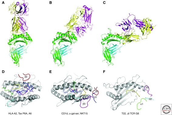
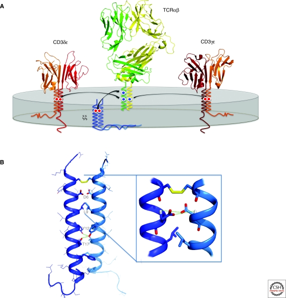
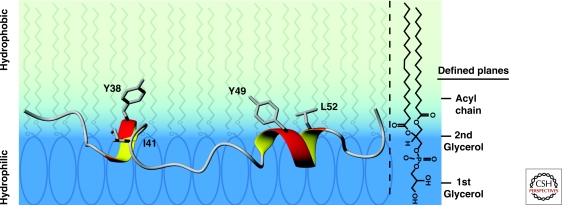
The T-cell receptor (TCR)-CD3 complex serves as a central paradigm for general principles of receptor assembly, ligand recognition, and signaling in the immune system. There is no other receptor system that matches the diversity of both receptor and ligand components. The recent expansion of the immunological structural database is beginning to identify key principles of MHC and peptide recognition. The multicomponent assembly of the TCR complex illustrates general principles used by many receptors in the immune system, which rely on basic and acidic transmembrane residues to guide assembly. The intrinsic binding of the cytoplasmic domains of the CD3epsilon and zeta chains to the inner leaflet of the plasma membrane represents a novel mechanism for control of receptor activation: Insertion of critical CD3epsilon tyrosines into the hydrophobic membrane core prevents their phosphorylation before receptor engagement.


Figures

Figure 1.
Figure 1.
TCR structures with different classes of ligands show distinct binding solutions. (A–C). The docking topologies are compared for the αβTCR A6 bound to a peptide-MHC class I complex (HLA-A2 with Tax P6A peptide; PDB entry 1QRN) (A), the αβTCR NKT15 bound to the complex of CD1d and the glycolipid α-galactosylceramide (PDB entry 2PO6) (B), and the γδTCR G8 bound to the nonclassical MHC class Ib molecule T22 (PDB entry 1YPZ) (C). (D–F) The placement of the TCR loops is compared for the same complexes. (D) The A6 TCR uses all six loops to contact the MHC molecule and the bound peptide. The four germline-encoded loops (α1, α2, β1, and β2) contribute to MHC binding, while the two CDR3 loops are positioned over the peptide (α3 and β3). (E) The NKT15 TCR contacts the CD1d-bound glycolipid only through germline elements encoded in the invariant TCRα chain (α1 and α3 loops). (F) The γδ TCR G8 inserts the CDR3δ chain loop into the hydrophobic groove of T22; other TCR loops do not appear to be essential for T22 binding.
Figure 2.
Figure 2.
Conserved helical residues of MHC class I and class II proteins are often contacted by CDR1 and CDR2 residues of TCRs. Conserved MHC helical residues are highlighted in green for structures of H-2Kb (A) and I-Ab (B). The BM3.3 TCR (H-2Kb; PDB entry 1NAM) and the B3K506 TCR (I-Ab; PDB entry 3C5Z) contact these conserved helical MHC residues. TCR CDR1β and CDR2β are colored red, CDR3α and CDR3β orange, and CDR1α and CDR2α blue. Peptide residues are colored yellow. Colored cyan are the H2-Kb α1 and IAb α1 domains, and magenta H2-Kb α2 and IAb β1 domains.
Figure 3.
Figure 3.
Assembly of the TCR-CD3 complex. (A) The TCR-CD3 complex is composed of four dimeric modules: TCRαβ (or γδ), CD3δε, CD3γε, and ζζ, which associate through intramembrane contacts to form the intact complex. Each dimeric signaling module associates with the TCR through a pair of acidic TM residues that bind a specific basic TM residue in the TCR. The result is an octameric complex with the stoichiometry as shown. Ribbon structures were generated from PDB entries 1MI5 (LC13 αβTCR), 1XMW (CD3δε), and 1SY6 (CD3γε). (B) NMR structure of the ζζ TM homodimer in detergent micelles (PBD entry 2HAC). The aspartic acid pair (D6-D6) that mediates assembly with TCR is located at the helix dimer interface and stabilized by interhelical H-bonds between side-chain oxygens and backbone amide protons (indicated by dotted lines). Further interface contacts include methyl and aromatic packing (such as L9-L9) and two tyrosine-threonine H-bonds (between Y12 and T17) that are critical for dimer formation.
Figure 4.
Figure 4.
Structure of the CD3ε ITAM in the lipid-bound state. The two tyrosines (Y38 and Y49) and the two aliphatic residues (I41 and L52) of the ITAM protrude into the hydrophobic acyl chain region of the lipid bilayer. The peptide backbone of the ITAM resides primarily at the interface between the hydrated lipid headgroup region and the hydrophobic bilayer interior. The hydrophobic layer of the bilayer is shaded light blue and the hydrated lipid headgroup region in dark blue. The POPG structure graphic to the right shows the location of the hydrophilic headgroup and the hydrophobic acyl chains of the lipid (PDB entry 2K4F).

References

    1. Adams EJ, Chien YH, Garcia KC 2005. Structure of a γδ T cell receptor in complex with the nonclassical MHC T22. Science 308:227–231 - PubMed
    1. Adams EJ, Strop P, Shin S, Chien YH, Garcia KC 2008. An autonomous CDR3δ is sufficient for recognition of the nonclassical MHC class I molecules T10 and T22 by γδ T cells. Nat Immunol 9:777–784 - PMC - PubMed
    1. Aivazian D, Stern LJ 2000. Phosphorylation of T cell receptor zeta is regulated by a lipid dependent folding transition. Nat Struct Biol 7:1023–1026 - PubMed
    1. Alcover A, Mariuzza RA, Ermonval M, Acuto O 1990. Lysine 271 in the transmembrane domain of the T-cell antigen receptor β chain is necessary for its assembly with the CD3 complex but not for α/β dimerization. J Biol Chem 265:4131–4135 - PubMed
    1. Arnett KL, Harrison SC, Wiley DC 2004. Crystal structure of a human CD3-ϵ/δ dimer in complex with a UCHT1 single-chain antibody fragment. Proc Natl Acad Sci 101:16268–16273 - PMC - PubMed

Publication types

MeSH terms

Substances

LinkOut - more resources