Review
. 2010 May;2(5):a001875.
doi: 10.1101/cshperspect.a001875. Epub 2010 Mar 31.

Axon guidance molecules in vascular patterning

Affiliations
Review

Axon guidance molecules in vascular patterning

Ralf H Adams et al. Cold Spring Harb Perspect Biol. 2010 May.

Abstract

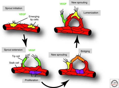
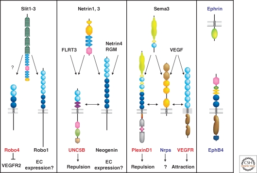
Endothelial cells (ECs) form extensive, highly branched and hierarchically organized tubular networks in vertebrates to ensure the proper distribution of molecular and cellular cargo in the vertebrate body. The growth of this vascular system during development, tissue repair or in disease conditions involves the sprouting, migration and proliferation of endothelial cells in a process termed angiogenesis. Surprisingly, specialized ECs, so-called tip cells, which lead and guide endothelial sprouts, share many feature with another guidance structure, the axonal growth cone. Tip cells are motile, invasive and extend numerous filopodial protrusions sensing growth factors, extracellular matrix and other attractive or repulsive cues in their tissue environment. Axonal growth cones and endothelial tip cells also respond to signals belonging to the same molecular families, such as Slits and Roundabouts, Netrins and UNC5 receptors, Semaphorins, Plexins and Neuropilins, and Eph receptors and ephrin ligands. Here we summarize fundamental principles of angiogenic growth, the selection and function of tip cells and the underlying regulation by guidance cues, the Notch pathway and vascular endothelial growth factor signaling.


Figures

Figure 1.
Figure 1.
Schematic organization of the blood vessel network. Blood flows (arrows) through arteries and arterioles into capillaries. In the embryonic dermis, arteries develop in close association with peripheral nerves, which are a source of VEGF. Venules and veins collect the blood from capillary beds. Vascular smooth muscle cells cover arteries and vein, whereas pericytes are confined to capillaries and postcapillary venules.
Figure 2.
Figure 2.
Angiogenic sprouting and blood vessel growth. ECs initiate sprouting in response to tissue-derived signals such as VEGF. A fraction of cells (shown in yellow and green) extends long filopodia and acquires motile and invasive behavior. These tip cells lead and guide new sprouts, whereas other ECs (shown in red) form the sprout stalk or stay behind to maintain tissue perfusion. At some point during sprout extension, tip cells will contact other tip cells or vessels to establish new connections. These cell bridges (orange) are converted into new blood-carrying vessels. Simultaneously, new sprouting is initiated at other sites (yellow and green cells) and additional ECs are generated by proliferation (purple).
Figure 3.
Figure 3.
Regulation of tip cell formation. Left: Image of sprouting ECs in the postnatal retina. New sprouting (yellow arrow), establish sprouts with distal tip cells (green arrows), ECs forming new connections (orange arrow), and perfused vessels (red asterisks) are highlighted. Right: Schematic showing the selection of a tip cell (green) through Notch signaling. High levels of Dll4 protein in tips activates Notch and dampens VEGF signaling in an adjacent stalk EC (red). Strong expression of Jagged1 in stalk cells antagonizes Dll4-mediated activation of Notch on neighboring tip ECs. Consequently, tip cells shown the strongest response to VEGF and grow toward the VEGF gradient. The levels of Notch signaling and the antagonistic activity of Jagged1 require Fringe-mediated glycosylation of Notch receptors.
Figure 4.
Figure 4.
Axon guidance receptor expression in ECs Schematic representation of the four families of axon guidance cues and their receptors. Predominantly endothelial-expressed receptors are labeled in red, receptors with shared expression in the nervous and the vascular system in blue and molecules with no (known) expression in the vascular system in black. Note that in each axon guidance receptor family, at least one member is expressed in the vasculature. See text for details.

References

    1. Adams RH, Alitalo K 2007. Molecular regulation of angiogenesis and lymphangiogenesis. Nat Rev Mol Cell Biol 8:464–478 - PubMed
    1. Adams RH, Wilkinson GA, Weiss C, Diella F, Gale NW, Deutsch U, Risau W, Klein R 1999. Roles of ephrinB ligands and EphB receptors in cardiovascular development: demarcation of arterial/venous domains, vascular morphogenesis, and sprouting angiogenesis. Genes Dev 13:295–306 - PMC - PubMed
    1. Appleton BA, Wu P, Maloney J, Yin J, Liang WC, Stawicki S, Mortara K, Bowman KK, Elliott JM, Desmarais W, et al.2007. Structural studies of neuropilin/antibody complexes provide insights into semaphorin and VEGF binding. EMBO J 26:4902–4912 - PMC - PubMed
    1. Armulik A, Abramsson A, Betsholtz C 2005. Endothelial/pericyte interactions. Circ Res 97:512–523 - PubMed
    1. Bagri A, Tessier-Lavigne M, Watts RJ 2009. Neuropilins in tumor biology. Clin Cancer Res 15:1860–1864 - PubMed

LinkOut - more resources