Review
. 2010 May;2(5):a002279.
doi: 10.1101/cshperspect.a002279.

ZAP-70: an essential kinase in T-cell signaling

Affiliations
Review

ZAP-70: an essential kinase in T-cell signaling

Haopeng Wang et al. Cold Spring Harb Perspect Biol. 2010 May.

Abstract

ZAP-70 is a cytoplasmic protein tyrosine kinase that plays a critical role in the events involved in initiating T-cell responses by the antigen receptor. Here we review the structure of ZAP-70, its regulation, its role in development and in disease. We also describe a model experimental system in which ZAP-70 function can be interrupted by a small chemical inhibitor.


Figures

Figure 1.
Figure 1.
A sequential model for T-cell activation. Following TCR engagement, CD4-associated Lck is brought into proximity of the CD3 complex and phosphorylates the ITAMs (phoshorylation depicted as red dots). Doubly phosphorylated ITAMs then interact with the tandem SH2 domains of ZAP-70. After ITAM binding, ZAP-70 can be phosphorylated by Lck, which results in activation of ZAP-70 catalytic activity and its autophosphorylation. Active ZAP-70 subsequently phosphorylates LAT and SLP-76, which function as scaffolds to recruit many other signaling molecules and lead to T-cell activation, proliferation, and differentiation (not shown).
Figure 2.
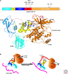
Figure 2.
Structural organization of autoinhibited ZAP-70, and model of autoinhibition. (A) Schematic of ZAP-70 domains and phosphorylated tyrosine residues with binding molecules (top) and a crystal structure of autoinhibited ZAP-70 (PDB code 2OZO, bottom). The amino-terminal SH2 domain, interdomain A, carboxy-terminal SH2 domain, interdomain B, and the kinase domain are shown in cyan, yellow, blue, red, and orange, respectively. The side chains of residues involved in the aromatic–aromatic interactions between interdomain A, interdomain B, and the kinase domain are labeled and colored in dark blue. Dotted lines represent disordered regions. (B) Model for the activation of ZAP-70 following ITAM binding. The SH2 domains, interdomain A, interdomain B, and kinase domains are colored as in (A). The doubly phosphorylated ITAM is depicted in pink. When ZAP-70 shows an autoinhibited conformation it is incompatible with binding to phosphorylated ITAMs. Following ITAM binding, conformational changes in ZAP-70 promote disassembly of the interface mediating the autoinhibited conformation, and exposure of tyrosines in interdomain B, leading to their phosphorylation that further destabilizes the interface.
Figure 3.
Figure 3.
Comparison of ZAP-70 with Syk. (A) Cartoon structures of the amino-terminal phosphotyrosine binding pocket in ZAP-70 (PDB code 2OQ1, left) and in Syk (PDB code 1A81, right). The amino-terminal SH2 domain, carboxy-terminal SH2 domain, and phosphorylated ITAM peptide are shown in cyan, blue, and pink, respectively. There are six different conformations in the crystal asymmetric unit of Syk. The Syk structure shown here is representative of three of six conformations. In the other three conformations, only one residue (Lysine) in the carboxy-terminal SH2 domain seems to interact with the phosphotyrosine of the ITAM. However, the electron density for the side-chain of this Lysine is absent in all three conformations (not shown), suggesting that this interaction may not contribute significantly to the binding. (B) Expression and function of Syk family kinases throughout T-cell development.
Figure 4.
Figure 4.
Mutations of ZAP-70 in Human SCID and hypomorphic mutant mice.
Figure 5.
Figure 5.
Chemical-genetic strategy to generate an inhibitor-sensitive ZAP-70. (A) Structure of the ZAP-70 kinase domain (PDB code 1U59). C- and N-lobes are indicated. The ATP-binding site is boxed. (B) Chemical structure of the inhibitor 3-MB-PP1. (C) Schematic of the analog-sensitive strategy. Left, Diagrams show the WT (top) and analog-sensitive mutant (bottom) ZAP-70 ATP-binding regions, modeled using PyMol. Circles identify the region of the gatekeeper residue. Right, Diagrams show the model of 3-MB-PP1 binding within the WT (top) and analog-sensitive mutant (bottom) ZAP-70. The presence of the “gatekeeper” residue in the ATP-binding site of WT ZAP-70 prevents the binding of the bulky 3-MB-PP1 inhibitor. In the analog-sensitive ZAP-70 mutant (bottom), the enlarged ATP-binding pocket allows binding of 3-MB-PP1. The 3-MB-PP1 model is derived from the crystal structure of the kinase domain of cryptosporidium parvum calcium dependent protein kinase in complex with 3-MB-PP1 (PDB code 2WEI). Top, white arrow points to the steric clash between 3-MB-PP1 and the gatekeeper residue, indicating that 3-MB-PP1 is not compatible with binding to WT ZAP-70.

References

    1. Allsup DJ, Kamiguti AS, Lin K, Sherrington PD, Matrai Z, Slupsky JR, Cawley JC, Zuzel M 2005. B-cell receptor translocation to lipid rafts and associated signaling differ between prognostically important subgroups of chronic lymphocytic leukemia. Cancer Res 65:7328–7337 - PubMed
    1. Arpaia E, Shahar M, Dadi H, Cohen A, Roifman CM 1994. Defective T cell receptor signaling and CD8+ thymic selection in humans lacking zap-70 kinase. Cell 76:947–958 - PubMed
    1. Ashe JM, Wiest DL, Abe R, Singer A 1999. ZAP-70 protein promotes tyrosine phosphorylation of T cell receptor signaling motifs (ITAMs) in immature CD4(+)8(+) thymocytes with limiting p56(lck). J Exp Med 189:1163–1168 - PMC - PubMed
    1. Au-Yeung BB, Deindl S, Hsu LY, Palacios EH, Levin SE, Kuriyan J, Weiss A 2009. The structure, regulation, and function of ZAP-70. Immunol Rev 228:41–57 - PubMed
    1. Bishop AC, Shah K, Liu Y, Witucki L, Kung C, Shokat KM 1998. Design of allele-specific inhibitors to probe protein kinase signaling. Curr Biol 8:257–266 - PubMed

Publication types

Substances