. 2010 Jul 16;285(29):22576-91.
doi: 10.1074/jbc.M110.118976. Epub 2010 May 7.

p62/SQSTM1 is a target gene for transcription factor NRF2 and creates a positive feedback loop by inducing antioxidant response element-driven gene transcription

Affiliations

p62/SQSTM1 is a target gene for transcription factor NRF2 and creates a positive feedback loop by inducing antioxidant response element-driven gene transcription

Ashish Jain et al. J Biol Chem. .

Abstract

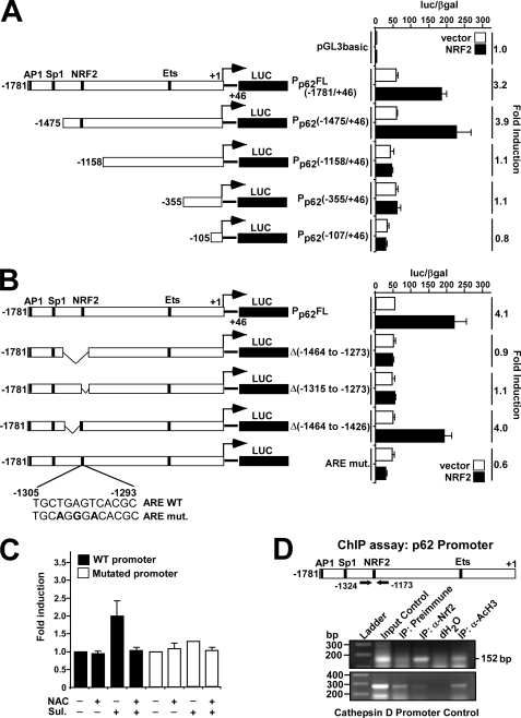
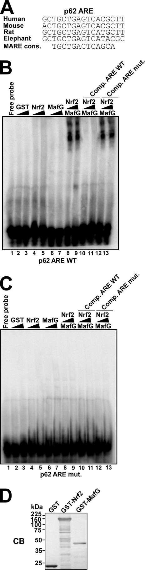
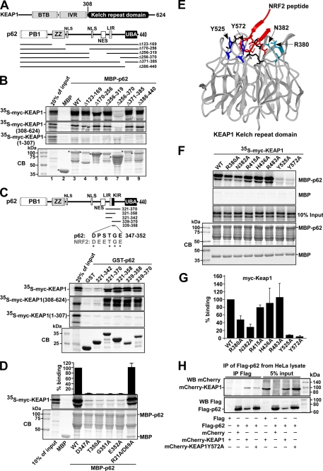
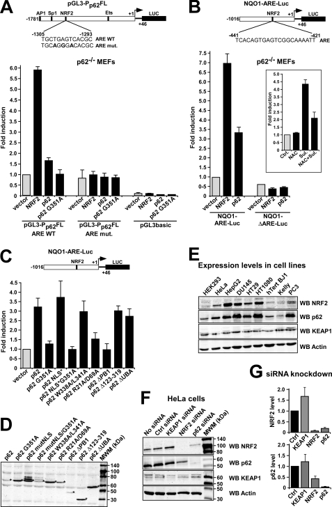
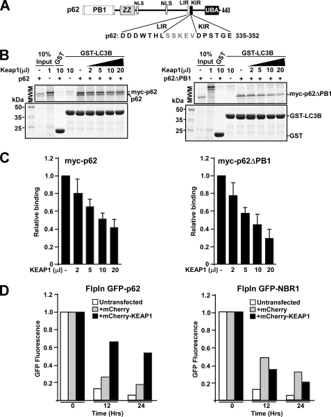
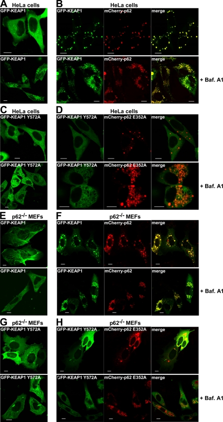
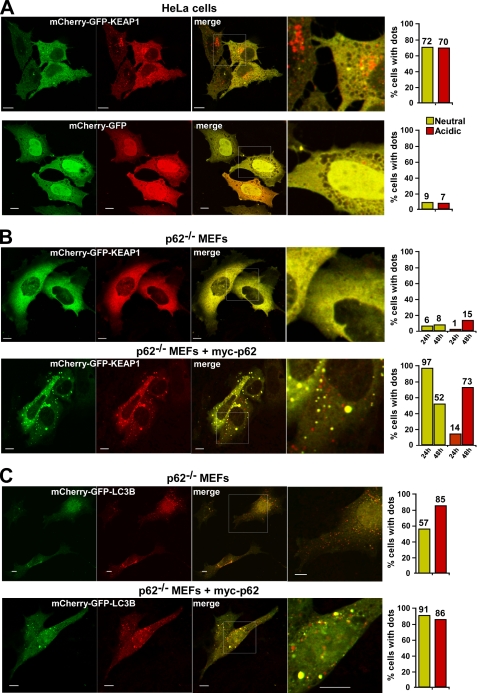
The p62/SQSTM1 (sequestosome 1) protein, which acts as a cargo receptor for autophagic degradation of ubiquitinated targets, is up-regulated by various stressors. Induction of the p62 gene by oxidative stress is mediated by NF-E2-related factor 2 (NRF2) and, at the same time, p62 protein contributes to the activation of NRF2, but hitherto the mechanisms involved were not known. Herein, we have mapped an antioxidant response element (ARE) in the p62 promoter that is responsible for its induction by oxidative stress via NRF2. Chromatin immunoprecipitation and gel mobility-shift assays verified that NRF2 binds to this cis-element in vivo and in vitro. Also, p62 docks directly onto the Kelch-repeat domain of Kelch-like ECH-associated protein 1 (KEAP1), via a motif designated the KEAP1 interacting region (KIR), thereby blocking binding between KEAP1 and NRF2 that leads to ubiquitylation and degradation of the transcription factor. The KIR motif in p62 is located immediately C-terminal to the LC3-interacting region (LIR) and resembles the ETGE motif utilized by NRF2 for its interaction with KEAP1. KIR is required for p62 to stabilize NRF2, and inhibition of KEAP1 by p62 occurs from a cytoplasmic location within the cell. The LIR and KIR motifs cannot be engaged simultaneously by LC3 and KEAP1, but because p62 is polymeric the interaction between KEAP1 and p62 leads to accumulation of KEAP1 in p62 bodies, which is followed by autophagic degradation of KEAP1. Our data explain how p62 contributes to activation of NRF2 target genes in response to oxidative stress through creating a positive feedback loop.


Figures

FIGURE 1.
FIGURE 1.
Mapping of an NRF2 binding site in the p62 promoter/enhancer. A and B, reporter gene assays were performed using wild-type (−1781/+46) or the indicated deleted or mutated p62 promoter/enhancer constructs. HEK293 cells were co-transfected with an empty vector (pcDNA3.1) (100 ng) or a plasmid encoding murine Nrf2 (pcDNA3.1-V5-mNrf2) (100 ng) together with the indicated p62 promoter constructs (60 ng). Cells were harvested 24 h after transfection. The relative promoter activities are expressed as the ratio between measured luciferase and β-galactosidase activities. The data shown are the mean activities obtained in one experiment performed in triplicate, and are representative of three or more independent experiments. For each p62 promoter construct, NRF2-mediated fold-activation is shown to the right. C, HEK293 cells co-transfected with wild-type or mutated p62 promoter constructs were analyzed as in A. The cells were treated for 20 h with sulforaphane (Sul, 15 μm) or N-acetylcysteine (NAC, 5 mm) as indicated. The relative activity of the wild-type promoter was set to one. The mean -fold activation obtained in three independent experiments performed in triplicate is presented. D, ChIP assays show that NRF2 can associate with the p62 promoter. Extracts from HeLa cells (1.5 × 10⁷ cells per antibody) were immunoprecipitated with preimmune serum, polyclonal anti-NRF2 antibody, or anti-acetylated histone H3 antibody as a positive control. Input control (1:50) was also included. PCR analyses of the immunoprecipitated chromatin were carried out using primers flanking the ARE (position −1324 and −1173, respectively) (upper panel). PCR analyses of the precipitated chromatin using primers aligning to position −3351 and −3069 of the cathepsin D promoter were used as a control.
FIGURE 2.
FIGURE 2.
NRF2-MAFG binds to the ARE in the p62 promoter in vitro. A, the ARE in the p62 promoter is conserved in human, mouse, rat, and elephant species, and constitutes a subclass of cis-elements that can be classified as MAREs (Maf recognition elements). B and C, gel mobility-shift assays demonstrating binding of NRF2-MAFG to the wild-type NRF2-responsive ARE element in the p62 promoter (B) but not to the mutated ARE site (C). GST, GST-NRF2, and GST-MAFG proteins, expressed and purified from E. coli, were incubated with [γ-32P]ATP-labeled oligonucleotides (position −1303/−1288) containing the wild-type ARE motif in the p62 promoter (B) or the mutated ARE motif (C). The amounts of recombinant proteins incubated with DNA were 5 μg or 10 μg of GST (lanes 1 and 2), 0.75 μg or 3.75 μg of GST-NRF2 (lanes 3 and 4), 0.5 or 2.5 μg of GST-MAFG (lanes 5 and 6), 0.5 μg of GST-MAFG or 1.5 μg (lanes 7, 9, and 11), or 3.75 μg (lanes 8, 10, and 12) of GST-NRF2. Competition experiments with cold oligonucleotides (1 μg) containing the wild-type ARE (lanes 11 and 12) or mutated ARE (lanes 13 and 14) designed from the p62 promoter show that binding is specific for the wild-type element. D, Coomassie staining of an SDS-PAGE gel showing the GST fusion proteins used in B and C.
FIGURE 3.
FIGURE 3.
The p62 protein interacts with KEAP1 via a short, conserved sequence motif. A, maps of KEAP1 and p62 indicating positions of domains and the deletion constructs employed in B to map the interaction between the two proteins. B, MBP pulldown assays showing that amino acids 321–370 in p62 are essential for binding to KEAP1, and that p62 interacts with the Kelch-repeat domain of KEAP1. The KEAP1 constructs that are depicted were in vitro translated in the presence of [35S]methionine, and tested in MBP pulldown assays for interaction with the indicated MBP-p62 constructs. The bottom gel panel shows a Coomassie Blue (CB)-stained SDS-polyacrylamide gel with the various MBP-p62 constructs used as input in the pulldown assays. Full-length proteins are indicated with asterisks. C, GST pulldown assays showing that amino acids 339–358 of p62 are sufficient for the interaction with KEAP1. The GST-p62 constructs, which contained the indicated peptides from p62, were tested in GST pulldown assays for interaction with the indicated Myc-tagged portions of KEAP1 produced by in vitro translation; full-length KEAP1 (1–624 amino acids), a C-terminal Kelch-repeat-containing fragment (308–624 amino acids), and an N-terminal BTB domain- and IVR-containing fragment (1–307 amino acids) were tested. The figure at the top shows the p62 constructs used and an alignment between p62 and NRF2 of the conserved DXXTGE motif (amino acids 347–352 in p62). D, MBP pulldown assays showing the effect of single point mutations in the DXXTGE motif of p62. Quantifications of the mean % binding with standard deviations from three independent experiments are shown above the autoradiograph. E, schematic based on the crystal structure of the Neh2 peptide from NRF2 bound to the Kelch-repeat domain of KEAP1. Seven amino acid residues located in loops of the Kelch-repeat were selected for mutational analyses to determine their effect on binding to p62. F and G, mapping of amino acid residues in the Kelch-repeat domain important for interaction with p62. The indicated single-point mutation constructs of KEAP1 were tested in MBP pulldown assays with MBP-p62. Quantification of the mean % binding with standard deviations from three independent experiments are shown. H, mCherry-KEAP1 co-immunoprecipitated with FLAG-p62 from HeLa cell extracts. Cells were co-transfected with the indicated constructs and FLAG-p62 immunoprecipitated with FLAG antibodies 24 h after transfection. Precipitated FLAG-p62 and co-precipitated mCherry-KEAP1 were detected by Western blotting using the indicated antibodies. For B, C, and H, data representative of three independent experiments with similar results are shown.
FIGURE 4.
FIGURE 4.
Reciprocal regulation of p62 and NFR2. A and B, ARE-element containing promoters are induced by p62. Reporter gene assays testing activation of the indicated p62 promoter (A) and Nqo1 promoter (B) reporter constructs by co-expression of Nrf2 or p62 in p62−/− MEFs. Cells were co-transfected with 100 ng of empty vector, V5-Nrf2, Myc-p62, or Myc-p62 G351A, together with 60 ng of the indicated Luciferase reporter constructs. The inset in B shows the effect on the activation of the Nqo1-ARE-Luc reporter of treating p62−/− MEFs with N-acetylcysteine (NAC) and sulforaphane (Sul) as indicated. C, reporter gene assays testing activation of the Nqo1-ARE-Luc reporter by co-expression of wild type and various mutants of p62 in p62−/− MEFs. Cells were co-transfected with the indicated Myc-p62 constructs together with 60 ng of Nqo1-ARE-Luc reporter. Cells were analyzed 24 h after transfection. The data in A, B, and C show the mean -fold induction with standard deviations based on three independent experiments performed in triplicate. D, Western blot experiment demonstrating expression of the various Myc-tagged p62 constructs in transfected p62−/− MEFs. The cell extracts were harvested 24 h post transfection using anti-Myc antibodies. E, the endogenous expression level of p62 correlates with the expression level of endogenous NRF2 in various human cell lines. Extracts of the indicated human cell lines were analyzed by Western blotting using the shown antibodies. The anti-actin blot shows equal amounts of proteins in the cell lysates loaded on the gel. F and G, knockdown of p62 in HeLa cells reduce the level of NRF2 and vice versa. HeLa cells transfected with KEAP1, NRF2, or p62 siRNAs, respectively, were analyzed by Western blotting using the indicated antibodies. Scrambled siRNA and mock transfection were used as controls. The quantifications are based on three independent experiments.
FIGURE 5.
FIGURE 5.
KEAP1 competes with LC3B for the interaction with p62. A, map of p62 illustrating the proximal location of the LIR (LC3-interacting region) and KIR (KEAP1 interacting region) motifs. B and C, GST pulldown assays demonstrating competition between LC3B and KEAP1 for binding to p62. GST-LC3B was incubated with in vitro translated wild-type p62 (polymeric) or a PB1 p62 deletion mutant (monomeric), in the presence or absence of increasing amounts of in vitro translated KEAP1. The data show that with increasing concentrations of KEAP1 there is a reduction in the amount of p62 bound to LC3B. The amount of p62 and LC3B is constant in all reactions. D, KEAP1 inhibits autophagic degradation of GFP-p62, but not GFP-NBR1, in HEK293 Flp-In T-Rex cells stably expressing the GFP-tagged proteins from a tetracycline-inducible promoter. Cells were grown overnight in rich medium (10% serum) in the presence of tetracycline, resulting in the accumulation of the GFP-tagged protein. Then tetracycline was removed (promoter shut-off), and degradation of the GFP-tagged protein followed for 24 h. Degradation of the GFP-tagged protein was measured at indicated time points by flow cytometry, and the readout was a loss of green fluorescence. Cells that were analyzed were either untransfected or transiently transfected with mCherry or mCherry-KEAP1. Expression of mCherry or mCherry-KEAP1 was verified by flow cytometry.
FIGURE 6.
FIGURE 6.
Overexpressed KEAP1 is recruited to p62 bodies and degraded by autophagy. A–D, HeLa cells were transfected as indicated with wild-type or mutated GFP-KEAP1 (Y572A), either alone (A and C) or together with mCherry-p62 (B and D). Bafilomycin A1 (16 h) was added as indicated (lower panels). E–H, p62−/− MEFs were transfected as indicated with wild-type or mutated GFP-KEAP1 (Y572A), either alone (E and G) or together with mCherry-p62 (F and H). Bafilomycin A1 (16 h) was added as indicated. A–H, cells were analyzed by confocal microscopy 24 h after transfection. Bars, 10 μm.
FIGURE 7.
FIGURE 7.
Overexpressed KEAP1 accumulates in acidic vesicles. A, HeLa cells transfected with mCherry-GFP-KEAP1 or mCherry-GFP were analyzed by confocal microscopy 24 h after transfection. The fraction of cells with mCherry-GFP-KEAP1 in neutral dots (green and red) and acidic dots (only red) was counted, and the result of a representative experiment based on counting of >300 transfected cells is shown to the right. B, p62−/− MEFs transfected with mCherry-GFP-KEAP1, either alone or together with Myc-p62, were analyzed by confocal microscopy 24 and 48 h after transfection. C, p62−/− MEFs transfected with mCherry-GFP-LC3B, either alone or together with Myc-p62, were analyzed by confocal microscopy 24 h after transfection. To the right is shown quantifications of the percentage of cells with neutral or acidic dots for representative experiments, each based on counting of more than 150 transfected cells. In A–C, bars represent 10 μm.

Similar articles

Cited by

References

    1. Halliwell B., Gutteridge J. M. (eds) (2007) Free Radicals in Biology and Medicine, 4th Ed., pp. 187–267, Oxford University Press, Oxford, UK
    1. Hayes J. D., McMahon M. (2009) Trends Biochem. Sci. 34, 176–188 - PubMed
    1. Kensler T. W., Wakabayashi N., Biswal S. (2007) Annu. Rev. Pharmacol. Toxicol. 47, 89–116 - PubMed
    1. Motohashi H., Yamamoto M. (2004) Trends Mol. Med. 10, 549–557 - PubMed
    1. Nguyen T., Nioi P., Pickett C. B. (2009) J. Biol. Chem. 284, 13291–13295 - PMC - PubMed

Publication types

MeSH terms