. 2010 Jul 9;285(28):21636-43.
doi: 10.1074/jbc.M109.088005. Epub 2010 May 7.

Aberrant amyloid precursor protein (APP) processing in hereditary forms of Alzheimer disease caused by APP familial Alzheimer disease mutations can be rescued by mutations in the APP GxxxG motif

Affiliations

Aberrant amyloid precursor protein (APP) processing in hereditary forms of Alzheimer disease caused by APP familial Alzheimer disease mutations can be rescued by mutations in the APP GxxxG motif

Lisa-Marie Munter et al. J Biol Chem. .

Abstract

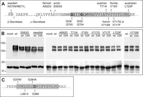
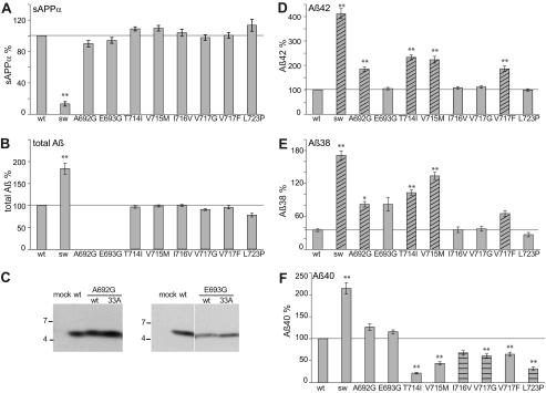
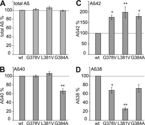
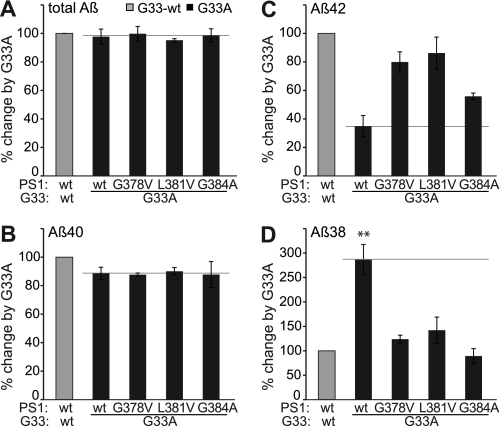
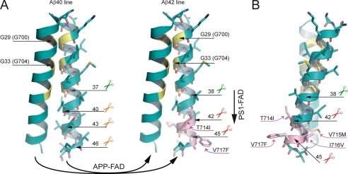
The identification of hereditary familial Alzheimer disease (FAD) mutations in the amyloid precursor protein (APP) and presenilin-1 (PS1) corroborated the causative role of amyloid-beta peptides with 42 amino acid residues (Abeta42) in the pathogenesis of AD. Although most FAD mutations are known to increase Abeta42 levels, mutations within the APP GxxxG motif are known to lower Abeta42 levels by attenuating transmembrane sequence dimerization. Here, we show that aberrant Abeta42 levels of FAD mutations can be rescued by GxxxG mutations. The combination of the APP-GxxxG mutation G33A with APP-FAD mutations yielded a constant 60% decrease of Abeta42 levels and a concomitant 3-fold increase of Abeta38 levels compared with the Gly(33) wild-type as determined by ELISA. In the presence of PS1-FAD mutations, the effects of G33A were attenuated, apparently attributable to a different mechanism of PS1-FAD mutants compared with APP-FAD mutants. Our results contribute to a general understanding of the mechanism how APP is processed by the gamma-secretase module and strongly emphasize the potential of the GxxxG motif in the prevention of sporadic AD as well as FAD.


Figures

FIGURE 1.
FIGURE 1.
APP- and PS1-FAD mutations. A, part of the APP sequence is shown. Indicated are the β- and α-secretase cleavage sites as well as all analyzed APP-FAD mutations. The glycine residues mediating the APP-TMS dimerization (9) are highlighted (Gly29/Gly700 and Gly33/Gly704 according to Aβ or APP770 numbering, respectively). B, APP-FAD and APP-FAD-G33A constructs are equally well and stably expressed in the SH-SY5Y cells. The double band represents mature, plasma membrane-residing (∼130 kDa) and immature (∼110 kDa) APP, and Western blot was stained with 22C11. As loading control, actin was stained as shown in the lower panel (∼40 kDa). C, an amino acid sequence of PS1 TMS-7 is shown. Analyzed FAD mutations and the catalytically active Asp385 are highlighted. The gray boxes in C mark residues embedded in the cell membrane.
FIGURE 2.
FIGURE 2.
Analysis of APP-FAD mutations. Quantification of secreted sAPPα, total Aβ, Aβ42, Aβ40, and Aβ38. A, sAPPα levels, APP-wt (without FAD) set as 100% (mean ± S.E., n = 5–16). B, total secreted Aβ levels, APP-wt set as 100% (mean ± S.E., n = 6–14). Note that A692G and E693G could not be analyzed by total Aβ ELISA as the FAD mutations alter the epitope of the antibody 4G8. C, total Aβ levels of A692G and E693G were analyzed by immunoprecipitation with antibody 18-1 and Western blot analysis with W0-2. D, Aβ42 levels, APP-wt set as 100% (mean ± S.E., n = 5–17). E, Aβ38 levels, APP-G33A set as 100%, see supplemental Fig. S1C and (mean ± S.E., n = 4–12). F, Aβ40 levels, APP-wt set as 100% (mean ± S.E., n = 7–16). A–E, asterisks indicate significant differences to APP-wt (*, p < 0.01, **, p < 0.001, one-way ANOVA type Dunnett). Shaded bars indicate mutations increasing Aβ42 and Aβ38. Horizontal shaded bars highlight mutations only decreasing Aβ40. Horizontal lines mark the fragment levels of APP-wt for better comparison with the mutants.
FIGURE 3.
FIGURE 3.
Impact of the APP-GxxxG mutation G33A on APP-FAD processing. A, Aβ42 levels; APP-wt was set as 100% (mean ± S.E., n = 5–17). For better comparison, bars of APP-FAD mutations as in Fig. 2D are shown (G33-wt, gray bars). The mutation G33A leads to a relative decrease in Aβ42 levels (black bars). Asterisks indicate significant differences to APP-wt (*, p < 0.01, **, p < 0.001, one-way ANOVA type Dunnett). The horizontal line marks Aβ42 level of APP-wt for better comparison. Original data of sAPPα, total Aβ, Aβ40, and Aβ38 ELISA are shown in supplemental Fig. S1. B–D, Aβ levels of the G33A constructs (black bars) normalized to the respective Gly33-wt constructs, which were set as 100% (represented by gray bars). Asterisks indicate significant differences to the respective Gly33-wt construct (**, p < 0.001, one-way ANOVA type Bonferroni). B, relative Aβ40 levels (mean ± S.E., n = 7–16). C, relative Aβ42 levels (mean ± S.E., n = 5–17). D, relative Aβ38 levels (mean ± S.E., n = 4–12). B–D, horizontal lines mark the fragment levels of APP-G33A for better comparison with the FAD mutants. E, analysis of expression levels of SPA4CT-related constructs in SH-SY5Y cells. The fragments show comparable expression levels, although these are generally lower than for SPA4CT-wt. Western blot was stained with monoclonal W0-2. F, levels of secreted Aβ from SPA4-fragments are normalized to SPA4CT-wt, which was set as 100% (mean ± S.E., n = 3–9). The mutation G33A has no impact on Aβ40 levels but decreases Aβ42 and increases Aβ38 levels. w/o, without.
FIGURE 4.
FIGURE 4.
Analysis of PS1-FAD mutants in the Gly33-wt background. Quantification of secreted Aβ from SH-SY5Y cells coexpressing PS1-FAD mutants and SPA4CT-wt. PS1-wt was set as 100%. A, total Aβ levels (mean ± S.E., n = 3–10). B, Aβ40 levels (mean ± S.E., n = 3–10). C, Aβ42 levels (mean ± S.E., n = 3–8). D, Aβ38 levels (mean ± S.E., n = 3–9). Asterisks indicate significant differences to PS1-wt/SPA4CT-wt (*, p < 0.01, **, p < 0,001, one-way ANOVA type Dunnett).
FIGURE 5.
FIGURE 5.
Impact of the GxxxG mutation G33A on PS1-FAD processing. ELISA data from PS1-FAD and SPA4CT-G33A coexpressing cells (black bars) in comparison to the respective SPA4CT-wt expressing lines, set as 100% (gray bars). A, total Aβ levels (mean ± S.E., n = 3–10). B, Aβ40 levels (mean ± S.E., n = 3–10). C, Aβ42 levels (mean ± S.E., n = 3–8). D, Aβ38 levels (mean ± S.E., n = 3–9). Asterisks indicate significant differences to PS1-wt/SPA4CT-wt (**, p < 0.001, one-way ANOVA type Bonferroni). Original data of total Aβ, Aβ42, Aβ40, and Aβ38 ELISA are shown in supplemental Fig. S4.
FIGURE 6.
FIGURE 6.
Model of the pathological mechanisms caused by APP-FAD and PS1-FAD mutations. Oxygen atoms are depicted in red, nitrogen atoms are depicted in blue, and sulfur atoms are colored dark yellow. The glycine residues of the interface are highlighted in yellow, and APP-FAD mutations are colored pink. Scissors indicate peptide bonds that are cleaved by the γ-secretase. A, dimeric wt substrates are degraded by the γ-secretase predominantly by the Aβ40 line. Left, corresponding peptide bonds are indicated and are located at the dimer interface. We propose that APP-FAD mutations cause a general shift between the two product lines so that the Aβ40 line is down-regulated, and the Aβ42 line becomes a major degradation pathway. Right, APP-FAD, peptide bonds of the Aβ42 line are indicated. Note, that amino acid side chains from FAD mutations do not reach into the dimer interface. PS1-FAD mutants seem to cause substrate flux inhibition leading to a retarded processing within the Aβ42 line causing increased Aβ42 and decreased Aβ38 levels (vertical arrow). The dimer crossing point mediated by Gly29 (Gly700) and Gly33 (Gly704) may cause a steric hindrance and inhibit the consecutive γ-secretase processing leading predominantly to Aβ40 (Aβ40 line) or to Aβ42 (Aβ42 line) (9, 10). Mechanistically, the effect of G33A occurs after the effects of APP-FAD mutations explaining why G33A causes a constant reduction by 60% for Aβ42 and a 3-fold increase of Aβ38 in the presence of all individual APP-FAD mutations analyzed. B, the APP-FAD-TMS dimer in side view for better illustration of the FAD-causing amino acid side chains. T714I, V715M, I716V, and V717F stick out of the dimer interface and thus likely affect processing by modulating the substrate-enzyme recognition.

References

    1. Chen C. D., Oh S. Y., Hinman J. D., Abraham C. R. (2006) J. Neurochem. 97, 30–43 - PubMed
    1. Gralle M., Botelho M. G., Wouters F. S. (2009) J. Biol. Chem. 284, 15016–15025 - PMC - PubMed
    1. Kaden D., Munter L. M., Joshi M., Treiber C., Weise C., Bethge T., Voigt P., Schaefer M., Beyermann M., Reif B., Multhaup G. (2008) J. Biol. Chem. 283, 7271–7279 - PubMed
    1. Rossjohn J., Cappai R., Feil S. C., Henry A., McKinstry W. J., Galatis D., Hesse L., Multhaup G., Beyreuther K., Masters C. L., Parker M. W. (1999) Nat. Struct. Biol. 6, 327–331 - PubMed
    1. Scheuermann S., Hambsch B., Hesse L., Stumm J., Schmidt C., Beher D., Bayer T. A., Beyreuther K., Multhaup G. (2001) J. Biol. Chem. 276, 33923–33929 - PubMed

Publication types

MeSH terms