. 2010 Jul 9;285(28):21382-90.
doi: 10.1074/jbc.M110.115022. Epub 2010 May 7.

TRIF modulates TLR5-dependent responses by inducing proteolytic degradation of TLR5

Affiliations

TRIF modulates TLR5-dependent responses by inducing proteolytic degradation of TLR5

Yoon Jeong Choi et al. J Biol Chem. .

Abstract

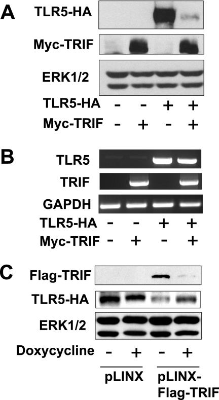
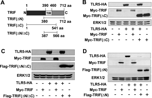
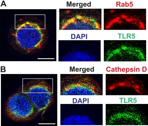
Proteolytic modification of pattern recognition receptors and their signaling adaptor molecules has recently emerged as an essential cellular event to regulate immune and inflammatory responses. Here we show that the TIR domain containing adaptor-inducing interferon-beta (TRIF), an adaptor molecule mediating TLR3 signaling and MyD88-independent signaling of TLR4, plays an inhibitory role in TLR5-elicited responses by inducing proteolytic degradation of TLR5. TRIF overexpression in human embryonic kidney (HEK293) and human colonic epithelial (NCM460) cells abolishes the cellular protein level of TLR5, whereas it does not alter TLR5 mRNA level. Thus, TRIF overexpression dramatically suppresses flagellin/TLR5-deriven NFkappaB activation in NCM460 cells. TRIF-induced TLR5 protein degradation is completely inhibited in the presence of pan-caspase inhibitor (benzyloxycarbonyl-Val-Ala-Asp-fluoromethyl ketone), whereas several specific inhibitors against cathepsin B, reactive oxygen species, or ubiquitin-mediated proteasome activity fail to suppress this degradation. These results indicate that TRIF-induced caspase activity causes TLR5 protein degradation. In addition, we identify that the C terminus of TRIF and extracellular domain of TLR5 are required for TRIF-induced TLR5 degradation. Furthermore, TRIF-induced proteolytic degradation is extended to TLR3, TLR6, TLR7, TLR8, TLR9, and TLR10, whereas the cellular level of TLR1, TLR2, and TLR4 is not affected by TRIF overexpression. These results suggest that, in addition to mediating TLR3- or TLR4-induced signaling as an adaptor molecule, TRIF can participate in proteolytic modification of certain members of TLRs to modulate the functionality of TLRs at post-translational level. Collectively, our findings propose a potential inhibitory role of TRIF at least in regulating host-microbial communication via TLR5 in colonic epithelial cells.


Figures

FIGURE 1.
FIGURE 1.
TNFα stimulation up-regulates TRIF expression in human colonic epithelial cells. A, total RNA was prepared from human colonic epithelial cells (NCM460) stimulated with TNFα (100 ng/ml) for 0, 4, and 8 h. Quantitative real-time PCR was performed to evaluate the mRNA expression level of TRIF, MyD88, TLR4, and TLR5. Error bars indicate S.D. B, NCM460 cells were stimulated with TNFα (100 ng/ml) for the indicated time periods. Lysates were immunoblotted using antibodies recognizing human TRIF or STAT3. C, the validity of human TRIF antibody used in this study was confirmed by immunoblot (IB) assay. Human TRIF transfected into HEK293 cells is evidently recognized by TRIF antibody. HEK293 cells were transfected with Myc-TRIF or empty vector. Lysates were immunoblotted using antibodies recognizing human TRIF, Myc, or STAT3. All data are representative of at least three independent experiments.
FIGURE 2.
FIGURE 2.
TRIF induces TLR5 protein degradation. A and B, HEK293 cells were co-transfected with TLR5-HA or Myc-TRIF. Lysates were immunoblotted using antibodies to HA, Myc, or ERK1/2 (A). TLR5 or TRIF gene expression was detected by PCR using primers: TLR5, 5′-TAT AAA GTC GAC GCC ACC ATG GGA GAC CAC CTG GAC-3′ and 5′-CTG ATG GCA TTG CTA AAG TTT CCT GTG-3′; TRIF, 5′-TTA TTA TTG CGG CCG CCA TGG CCT GCA CAG GCC CAT CAC TTC C-3′ and 5′-GGA TGT TTC TGG GGT GGT GGG AGT AGG-3′ (B). C, NCM460-TLR5-HA cells were transduced with retroviral particles harboring the doxycycline-inducible pLINX-FLAG-TRIF or the empty pLINX vector. The presence of doxycycline suppresses FLAG-TRIF expression. All data are representative of at least three independent experiments.
FIGURE 3.
FIGURE 3.
TRIF overexpression abolishes flagellin/TLR5-induced NFκB activation in human colonic epithelial cells (NCM460). A and B, NCM460 cells were co-transfected with NFκB-luciferase reporter and TRIF (A) or MyD88 (B) expression constructs or its empty vector as indicated. Luciferase activity was measured in the cells stimulated with flagellin (0, 50, 100, 200 ng/ml) for 6 h. Error bars indicate S.D. All data are representative of three independent experiments. All data are representative of at least three independent experiments. RLA, relative luciferase activity.
FIGURE 4.
FIGURE 4.
TRIF overexpression induces the protein degradation of various TLRs, except TLR1, TLR2, and TLR4. A–J, Myc-TRIF (4 μg) or FLAG-MyD88 (4 μg) construct was transiently co-transfected into HEK293 cells together with various human TLR expression constructs (2 μg) (TLR3 (A), TLR4 (B and C), TLR1 (D), TLR2 (E), TLR6 (F), TLR7 (G), TLR8 (H), TLR9 (I), and TLR10 (J)) as indicated. Total cell extracts were immunoblotted with HA, Myc, or FLAG antibody. ERK1/2 was determined as a loading control. All data are representative of at least three independent experiments.
FIGURE 5.
FIGURE 5.
TRIF, rather than MyD88, plays a critical role in modulating the cellular abundance of TLR5 protein. A, HEK293 cells were transfected with TLR5-HA (2 μg) and FLAG-MyD88 (4 μg) constructs. Total cell extracts were immunoblotted. B, TLR5 or MyD88 mRNA expression was detected by PCR using primers; MyD88, 5′-CTT GAT GAC CCC CTA GGA CA-3′ and 5′-CTG TTG GAC ACC TGG AGA CA-3′. C and D, TLR5-HA (2 μg) and FLAG-TRIF or FLAG-MyD88 constructs were transfected into HEK293 cells in an indicated combination. Although different amounts of FLAG-TRIF or FLAG-MyD88 construct were included as indicated, the total amount of DNA per each transfection was kept constant by adding its empty vector. Total ERK1/2 was evaluated as a loading control. All data are representative of at least three independent experiments.
FIGURE 6.
FIGURE 6.
C-terminal domain of TRIF is responsible for TRIF-induced TLR5 degradation. A, schematic diagram of various TRIF mutant constructs are shown. aa, amino acids; C, C terminus; N, N terminus; TIR, toll-interleukin 1 receptor. B–D, the full-length TLR5-HA construct was co-transfected with Myc-TRIF wild type and C-terminal deleted TRIF (Myc-TRIF(ΔC)) (B) or both N- and C-terminal deleted TRIF (FLAG-TRIF(ΔN/ΔC)) (C) or N-terminal deleted (FLAG-TRIF(ΔN)) (D) in HEK293 cells. Data are from one representative of three independent experiments.
FIGURE 7.
FIGURE 7.
Extracellular domain of TLR5 is required for TRIF-induced TLR5 degradation. A, various TLR5 mutants construct are shown. ECD, extracellular domain; LRR, leucine-rich repeat; TM, transmembrane; aa, amino acids. B and C, full-length TLR5-HA construct was co-transfected with Myc-TRIF wild type and extracellular domain-deleted TLR5 (TLR5 (ΔECD)-HA) (B) or TIR domain-deleted TLR5 (TLR5(ΔTIR)-HA) (C) in HEK293 cells. Data are from one representative of three independent experiments.
FIGURE 8.
FIGURE 8.
TRIF-induced caspase activity mediates TLR5 protein degradation. A–E, HEK293 cells were co-transfected with TLR5-HA and Myc-TRIF and then treated overnight with cathepsin B inhibitor (A), N-acetylcysteine (NAC) (B), NH4Cl (C), MG132 (D), or benzyloxycarbonyl-Val-Ala-Asp-fluoromethyl ketone (E). Lysates were immunoblotted using antibodies to HA, Myc, or ERK1/2. In the lower part of panel D, RAW264.7 cells were pretreated with MG132 or vehicle for 45 min followed by LPS (0.1 μg/ml) stimulation for 15 min. Lysates were immunoblotted using antibodies against cleaved caspase-3 or -8 or PARP (E). Data are representative of three independent experiments.
FIGURE 9.
FIGURE 9.
TLR5 is localized to endolysosomes by flagellin stimulation. A and B, NCM460 cells were transfected with TLR5-HA and then stimulated with flagellin (100 ng/ml) for 30 min. Co-localization of TLR5-HA and Rab5 (A) or cathepsin D (B) was shown. 4,6-Diamidino-2-phenylindole (DAPI) staining represents the nucleus. The inset area in the merged image was enlarged on the right. Data are representative of at least three independent experiments, and more than 95% of the cells had similar staining patterns. Fluorescence microscopy of the control cells stimulated with the vehicle was presented in supplemental Fig. 1.

Similar articles

Cited by

References

    1. Palm N. W., Medzhitov R. (2009) Immunol. Rev. 227, 221–233 - PubMed
    1. Medzhitov R., Preston-Hurlburt P., Kopp E., Stadlen A., Chen C., Ghosh S., Janeway C. A., Jr. (1998) Mol. Cell 2, 253–258 - PubMed
    1. Oshiumi H., Matsumoto M., Funami K., Akazawa T., Seya T. (2003) Nat. Immunol. 4, 161–167 - PubMed
    1. Kawai T., Akira S. (2008) Ann. N.Y. Acad. Sci. 1143, 1–20 - PubMed
    1. Kenny E. F., O'Neill L. A. (2008) Cytokine 43, 342–349 - PubMed

Publication types

LinkOut - more resources