. 2010 Sep;38(17):5867-83.
doi: 10.1093/nar/gkq349. Epub 2010 May 7.

Rapid decay of unstable Leishmania mRNAs bearing a conserved retroposon signature 3'-UTR motif is initiated by a site-specific endonucleolytic cleavage without prior deadenylation

Affiliations

Rapid decay of unstable Leishmania mRNAs bearing a conserved retroposon signature 3'-UTR motif is initiated by a site-specific endonucleolytic cleavage without prior deadenylation

Michaela Müller et al. Nucleic Acids Res. 2010 Sep.

Abstract

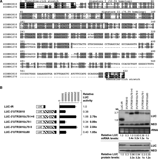
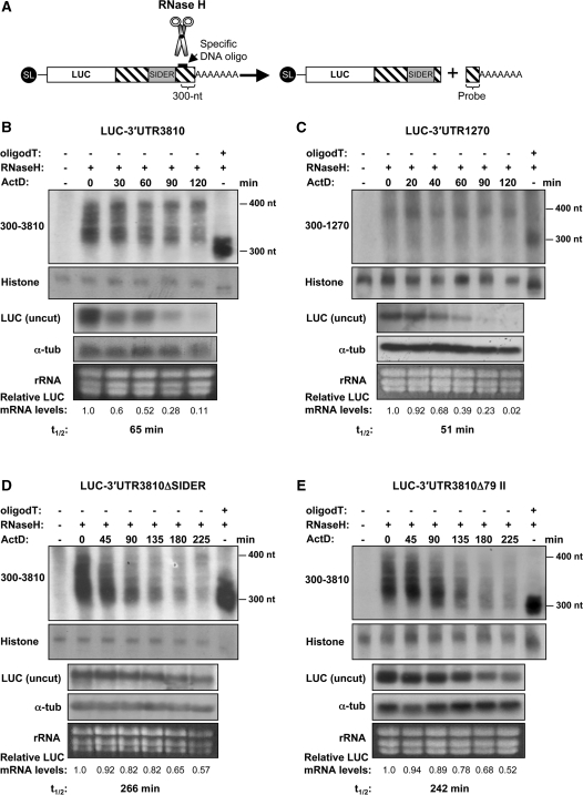
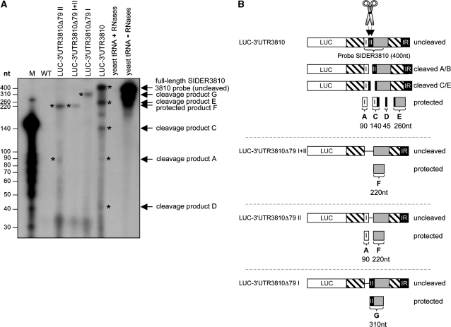
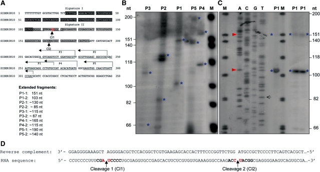
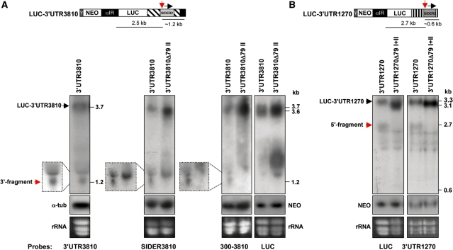
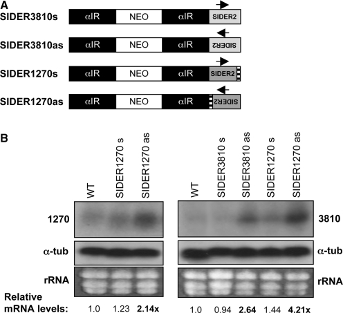
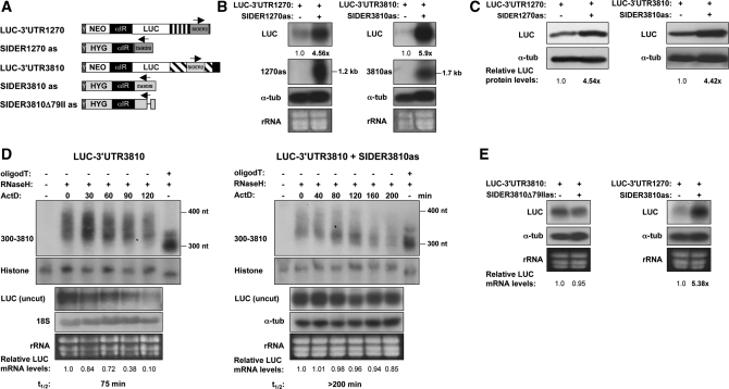
We have previously shown that the Leishmania genome possess two widespread families of extinct retroposons termed Short Interspersed DEgenerated Retroposons (SIDER1/2) that play a role in post-transcriptional regulation. Moreover, we have demonstrated that SIDER2 retroposons promote mRNA degradation. Here we provide new insights into the mechanism by which unstable Leishmania mRNAs harboring a SIDER2 retroposon in their 3'-untranslated region are degraded. We show that, unlike most eukaryotic transcripts, SIDER2-bearing mRNAs do not undergo poly(A) tail shortening prior to rapid turnover, but instead, they are targeted for degradation by a site-specific endonucleolytic cleavage. The main cleavage site was mapped in two randomly selected SIDER2-containing mRNAs in vivo between an AU dinucleotide at the 5'-end of the second 79-nt signature (signature II), which represents the most conserved sequence amongst SIDER2 retroposons. Deletion of signature II abolished endonucleolytic cleavage and deadenylation-independent decay and increased mRNA stability. Interestingly, we show that overexpression of SIDER2 anti-sense RNA can increase sense transcript abundance and stability, and that complementarity to the cleavage region is required for protecting SIDER2-containing transcripts from degradation. These results establish a new paradigm for how unstable mRNAs are degraded in Leishmania and could serve as the basis for a better understanding of mRNA decay pathways in general.


Figures

Figure 1.
Figure 1.
The second highly conserved 79-nt signature sequence amongst SIDER2 retroposons is essential for mRNA degradation. (A) Sequence alignment of two selected SIDER2 sequences present in the 3′-UTRs of L. major LmjF36.3810 (3810) and LmjF08.1270 (1270) transcripts using the multiple sequence alignment program multialin (http://bioinfo.genotoul.fr/multalin/multalin.html). Conserved motifs within SIDER2 retroposons, including the thymidine-rich stretch, the two tandemly repeated signatures I and II at the 5′-end of SIDER2, and the adenosine-rich stretch at the 3′-end are highlighted. (B) Schematic representation of the chimeric luciferase (LUC) constructs tested and their corresponding names. The full-length 3′-UTR of 3810 (3′-UTR3810), a 3′-UTR lacking both conserved signatures (3′-UTR3810Δ79I+II), a 3′-UTR lacking either the first (3′-UTR3810Δ79I) or the second (3′-UTR3810Δ79II) signature and a 3′-UTR lacking the last 80 nt of SIDER3810 (3′-UTR3810Δ80) were placed downstream of the LUC reporter gene. The entire SIDER2 element or its truncated forms are illustrated as grey boxes. The plasmid LUC-IR, containing the IR of 3810 served as a control. LUC activity was measured in the different transfectants (middle panel). Fold differences in LUC activity were normalized with plasmid copy numbers (Supplementary Table S2) and the values indicated are relative to the LUC-IR control. Numbers in bold correspond to the relative fold changes compared to the full-length 3′-UTR3810. Each bar and value represents the mean and standard deviations of four independent experiments. Northern and western blot analyses of total RNA and protein extracts from recombinant L. major promastigotes were carried out using LUC-specific probes and antibodies (right panels). Expression levels (mRNA and protein) of the NEO marker present on all plasmids served as a control for loading and for determining differences in plasmid copy numbers. Signal intensities were quantified and normalized with respect to the loading controls and plasmid copy numbers.
Figure 2.
Figure 2.
Rapid degradation of unstable SIDER2-bearing transcripts is not initiated by a shortening of the poly(A) tail. (A) Schematic representation of the deadenylation assay. LUC transcripts are specifically cleaved at 300 nt from the poly(A) tail using oligonucleotide-directed RNase H cleavage. The resulting 3′-products containing the poly(A) tail are visualized by northern blot using a probe complementary to the last 300 nt of transcripts 3810 and 1270, respectively. Poly(A) tail lengths of chimeric LUC transcripts were analyzed at different time points after transcriptional arrest using ActD. In each experiment, one sample was treated with oligo(dT) and served as a control for a completely deadenylated mRNA species. Another RNA sample that was not treated with RNase H was used as negative control. Deadenylation profiles of the unstable SIDER2-containing transcripts (B) LUC-3′-UTR3810 and (C) LUC-3′-UTR1270 and of stable LUC chimeric mRNAs lacking either the complete (D) SIDER3810 or (E) signature II. Histone 4A mRNA was used as a loading control together with an ethidium bromide staining to visualize rRNA. Decay kinetics of the corresponding uncut LUC mRNAs (from identical samples) and their half-lives (t1/2) are shown below the blots to demonstrate the fate of the full-length LUC transcripts (uncut) in comparison to the cleaved 3′-ends including the poly(A) tails. LUC mRNA levels were normalized to those of the α-tubulin mRNA. The numbers indicated below the blots represent the relative LUC transcript abundance with respect to time point 0 (before addition of the transcription inhibitor Act D). Deadenylation assays shown here are representative of three independent experiments that yielded similar results.
Figure 3.
Figure 3.
Detection of in vivo-generated cleavage products derived from SIDER2-containing mRNAs by RPAs. (A) Total RNA was isolated from L. major wild-type cells (WT), the recombinant L. major expressing a chimeric LUC transcript carrying the full-length 3′-UTR of 3810 (LUC-3′-UTR3810) and the truncated mutants LUC-3′-UTR3810Δ79I+II, LUC-3′-UTR3810Δ79I and LUC-3′-UTR3810Δ79II lacking either both signatures (I+II) or only signature I or signature II, respectively. These RNAs were independently mixed with an in vitro-transcribed radiolabeled anti-sense SIDER3810 probe of 400 nt and thereafter subjected to RNase A/T1 treatment. Wild-type RNA and yeast RNA with (negative control) or without RNase treatment (positive control) were used as controls. A 3′-labeled RNA ladder was used to estimate the size of the fragments (M). (B) Schematic representation of the in vivo cleaved (unstable LUC-3′-UTR3810 and LUC-3′-UTR3810Δ79I) and uncleaved (stabilized LUC-3′-UTR3810Δ79I+II and LUC-3′-UTR3810Δ79II) SIDER2-containing mRNAs. The sizes of the observed cleavage products suggest that a first cleavage occurs at the beginning of SIDER3810 signature II (cleavage A/B) and that a second, more dominant, cleavage occurs at ∼50-nt downstream (cleavage C/E). In addition to the full-length protected band (400 nt), four cleavage products of ∼260, ∼140, ∼90 and ∼45 nt (indicated with asterisks) were detected from the LUC-3′-UTR3810 mRNA. The cleavage products of 260 and 140 nt correspond to the second cleavage and the 90-nt fragment to the first cleavage (the expected 310-nt fragment is not visible in this experiment, as it was further cleaved at the second position to generate the 45- and 260-nt fragments). The band observed between the cleavage fragments C and E in the LUC-3′-UTR3810 lane was not reproducible in the other experiments. In all cases, cleavage products sum up to the size of the full-length SIDER3810 probe (400 nt). In the truncated LUC-3′-UTR3810Δ79I mRNA-lacking signature I, only the first cleavage site was detected, as a single fragment of ∼310 nt was protected. In the case of LUC-3′-UTR3810Δ79II mRNA-lacking signature II, two cleavage fragments of 90 and 220 nt were detected, as expected, given that RNases could digest the ss region of the probe corresponding to signature II. Deletion of both signatures in LUC-3′-UTR3810Δ79I+II mRNA produced a 220-nt RNase-resistant fragment, as expected. The data shown here are representative of three independent experiments that generated the same RNase digestion patterns.
Figure 4.
Figure 4.
Mapping of the cleavage site(s) in a SIDER2-containing mRNA by primer extension analysis. (A) Nucleotide sequence of SIDER3810 retroposon with signature I (black box) and II (grey box) sequences highlighted, and the five primers (P1–P5) indicated by arrows. The sizes of the expected primer extension products with full-length LUC-3′-UTR3810 mRNA, depending on whether cleavage occurs at the first (Cl1) or the second (Cl2) site within signature II, are listed below. (B) Primer extension assay with total RNA isolated from L. major LUC-3′-UTR3810 transfectant using five different reverse primers P1–P5 (Supplementary Table S1). Two specific extension fragments were obtained with each of the primers (indicated with blue asterisks) corresponding to both cleavages (1 and 2). Their sizes estimated with a radiolabeled DNA marker (M) are in agreement with the predicted cleavage regions. (C) A dideoxy-sequencing and primer extension with primer P1 allowed a precise mapping of the cleavage sites between an AU dinucleotide (cleavage 1) and a CU (or a UA; it is not easy to distinguish from the sequencing data) dinucleotide (cleavage 2) marked with a red arrow. (D) Reverse complement sequence (the black arrow indicates where the highlighted sequence starts) and the corresponding sequence of SIDER3810 RNA. Cleavage sites 1 and 2 are indicated with an arrow. The data shown here are representative of four independent experiments with similar results.
Figure 5.
Figure 5.
Detection of in vivo-generated endonucleolytic cleavage products from reporter mRNAs harboring distinct SIDER2 retroposon elements by northern blotting. (A) Northern blots of total RNA from recombinant L. major LUC-3′-UTR3810 (LUC reporter gene under the control of the full-length 3′-UTR of transcripts 3810) and LUC-3′-UTR3810Δ79II (3′-UTR of 3810 lacking the SIDER2 signature II sequence) hybridized with four different DNA probes corresponding to the complete 3′-UTR3810 (left panel), the SIDER3810 sequence alone, the last 300 nt of the 3810 3′-UTR (middle panels) or the LUC gene (right panel). The full-length 3′-UTR3810, SIDER3810 and 300–3810 probes recognized a band of ∼1.2 kb, which corresponds to the expected 3′-cleavage product (indicated by a red arrow; see also schematic representation above the blots). This band is absent in the control RNA-lacking signature II (cleavage region). The 5′-cleavage fragment was not detected under these conditions using either the full-length 3′-UTR3810 or the LUC-specific probes. (B) Northern blots of total RNA from L. major transfectants LUC-3′-UTR1270 (LUC reporter gene under the control of the full-length 3′-UTR of transcripts 1270) and LUC-3′-UTR1270Δ79I+II (3′-UTR of 1270 lacking both signatures I and II) hybridized with two different DNA probes specific for LUC (left panel) and the 3′-UTR1270 (right panel). Both probes detected a band of ∼2.7 kb corresponding to the expected size of the 5′-cleavage product (indicated by a red arrow; see schematic representation above the blots), which is absent in the control RNA-lacking signatures I+II (cleavage region). The 3′-cleavage fragment was not detected under these conditions. Northern blots shown here are representative of three independent experiments yielding similar results. Expression levels of the NEO mRNA present on all plasmids served as a control for loading and for evaluating differences in plasmid copy numbers between the transfectants.
Figure 6.
Figure 6.
Ectopic expression of SIDER2 anti-sense RNA results in an increased accumulation of endogenous L. major SIDER2-containing transcripts. (A) Schematic representation of plasmids overexpressing SIDER2 in both orientations. SIDER2 sequences from transcripts 1270 or 3810 were cloned downstream of a NEO selection marker in the sense (s) or anti-sense (as) orientation and stably transfected into L. major wild-type cells (WT). αIR refers to the IR of the α-tubulin gene necessary for NEO mRNA processing. (B) Northern blot analysis with total RNA from L. major WT and transfectants. The endogenous transcripts 1270 or 3810 were detected with probes specific to the coding regions of 1270 (left panel) and 3810 (right panel). The copy number of the SIDER2 expression plasmids was estimated by Southern blot as indicated in Supplementary Table S2. The signal intensities were quantified and the mRNA abundance was normalized to α-tubulin mRNA and calculated with respect to WT mRNA levels.
Figure 7.
Figure 7.
SIDER2 anti-sense RNA complementary to the cleavage region blocks degradation of unstable SIDER2-containing reporter transcripts. (A) Schematic representation of the chimeric LUC reporter constructs bearing either a sense (s) or anti-sense (as) SIDER2 retroposon from transcripts 1270 or 3810. Pairs of sense/anti-sense vectors LUC-3′-UTR1270/SIDER1270as LUC-3′-UTR3810/SIDER3810as, LUC-3′-UTR3810/SIDER3810Δ79IIas and 3′-UTR1270/SIDER3810as were co-transfected into L. major. The plasmids expressing the full-length 3′-UTRs of 3810 and 1270 cloned downstream of the LUC gene harbor a neomycin phosphotransferase (NEO) gene as a selection marker. Anti-sense SIDER2 sequences were inserted downstream of a hygromycin B (HYG) selection marker. Arrows indicate the orientation of SIDER2 elements. Y corresponds to a 92-bp polypyrimidine stretch (34) and α-IR refers to the IR of α-tubulin, both necessary for NEO, HYG and SIDER2as RNA processing. (B) Northern blot analysis to compare LUC mRNA expression levels in single (−) and double (+) transfectants using a LUC-specific probe. Expression and correct processing of both fully complementary anti-sense SIDER2 transcripts (1270as and 3810as) was verified with riboprobes specific to each SIDER2 sequence. Their sizes of ∼1.2 kb indicate that the transcripts are spliced within the α-IR and do not contain HYG sequences. The LUC signal intensities were normalized with α-tubulin mRNA levels and calculated with respect to the LUC-plasmid copy numbers (Supplementary Table S2). (C) Western blot analysis using L. major protein lysates and a LUC-specific antibody. Protein loading was controlled with an anti-α-tubulin antibody. (D) Deadenylation profile and decay kinetics in single (LUC-3′-UTR3810) and double L. major transfectants (LUC-3′-UTR3810/SIDER3810as) as determined by northern blotting (identical samples). The numbers below the blots represent the relative LUC mRNA levels with respect to time point 0 (before addition of ActD). Histone 4A was used as a loading control. The mRNA half-lives of the uncut LUC transcripts are shown below the blots. (E) Northern blot hybridization to evaluate LUC-3′-UTR3810 mRNA accumulation in the absence or presence of a truncated SIDER3810 anti-sense RNA-lacking signature II (SIDER3810Δ79IIas) (left panel). Northern blot analysis to compare LUC-3′-UTR1270 mRNA abundance in the presence or absence of a heterologous SIDER3810 anti-sense RNA (right panel). SIDER3810 and SIDER1270 retroposons share a 76.5% sequence identity at the level of signature II and >80% identity within the cleavage site (Figure 1A). LUC signal intensities were normalized as indicated in (B).

Similar articles

Cited by

References

    1. Myler PJ, Audleman L, deVos T, Hixson G, Kiser P, Lemley C, Magness C, Rickel E, Sisk E, Sunkin S, et al. Leishmania major Friedlin chromosome 1 has an unusual distribution of protein-coding genes. Proc. Natl Acad. Sci. USA. 1999;96:2902–2906. - PMC - PubMed
    1. Martinez-Calvillo S, Yan S, Nguyen D, Fox M, Stuart K, Myler PJ. Transcription of Leishmania major Friedlin chromosome 1 initiates in both directions within a single region. Mol. Cell. 2003;11:1291–1299. - PubMed
    1. Liang XH, Haritan A, Uliel S, Michaeli S. trans and cis splicing in trypanosomatids: mechanism, factors, and regulation. Eukaryot. Cell. 2003;2:830–840. - PMC - PubMed
    1. Clayton C, Shapira M. Post-transcriptional regulation of gene expression in trypanosomes and leishmanias. Mol. Biochem. Parasitol. 2007;156:93–101. - PubMed
    1. Haile S, Papadopoulou B. Developmental regulation of gene expression in trypanosomatid parasitic protozoa. Curr. Opin. Microbiol. 2007;10:569–577. - PubMed

Publication types