. 2010 Sep;38(17):5746-60.
doi: 10.1093/nar/gkq267. Epub 2010 May 7.

Aldosterone and vasopressin affect {alpha}- and {gamma}-ENaC mRNA translation

Affiliations

Aldosterone and vasopressin affect {alpha}- and {gamma}-ENaC mRNA translation

Andrea Perlewitz et al. Nucleic Acids Res. 2010 Sep.

Abstract

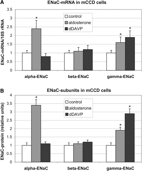
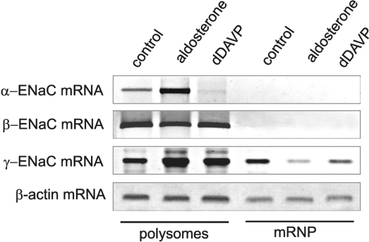
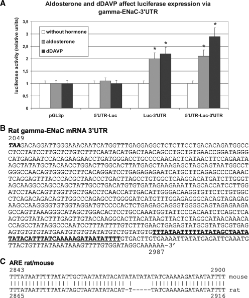
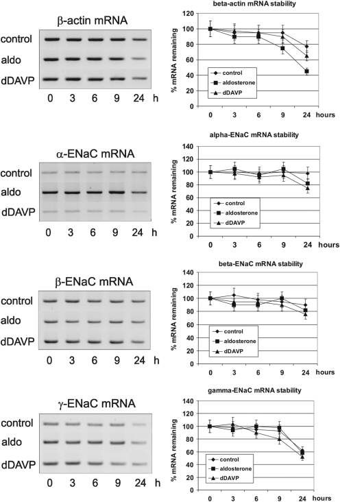
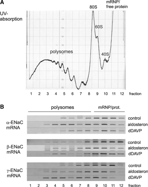
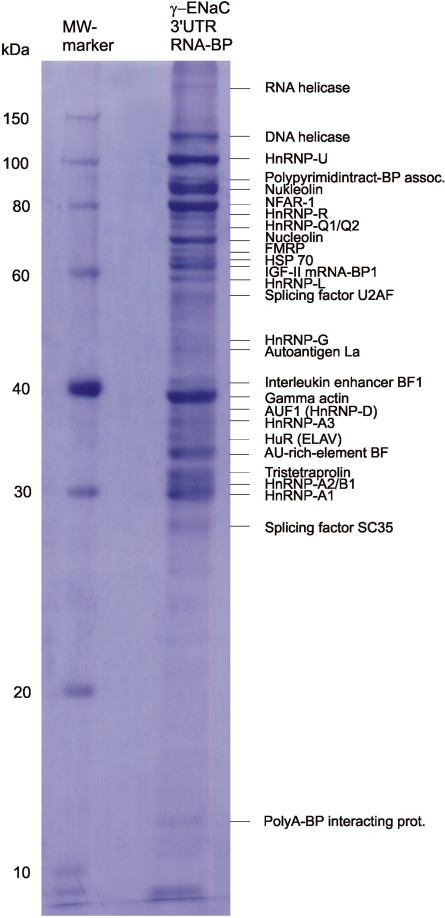
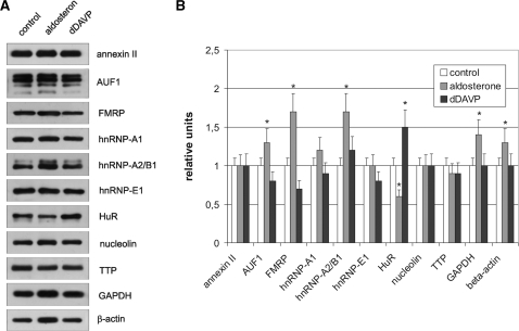
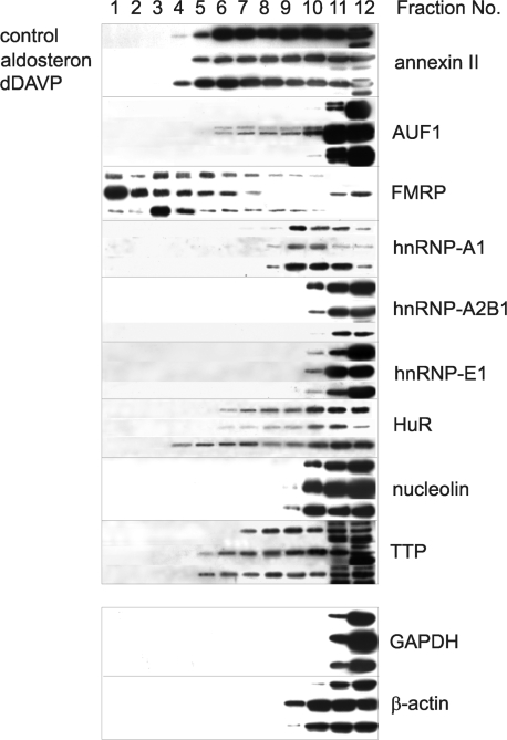
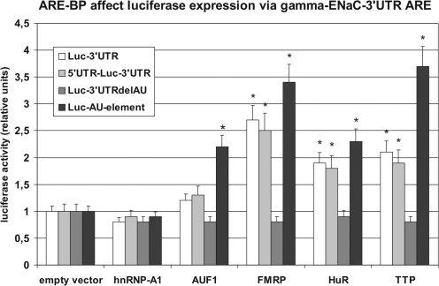
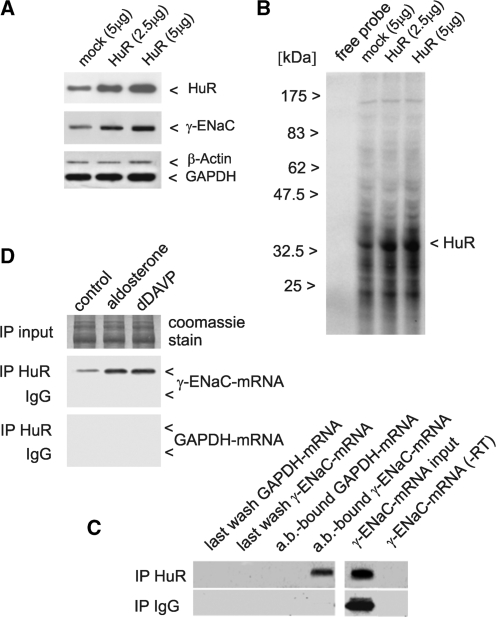
Vasopressin and aldosterone play key roles in the fine adjustment of sodium and water re-absorption in the nephron. The molecular target of this regulation is the epithelial sodium channel (ENaC) consisting of α-, β- and γ-subunits. We investigated mRNA-specific post-transcriptional mechanisms in hormone-dependent expression of ENaC subunits in mouse kidney cortical collecting duct cells. Transcription experiments and polysome gradient analysis demonstrate that both hormones act on transcription and translation. RNA-binding proteins (RBPs) and mRNA sequence motifs involved in translational control of γ-ENaC synthesis were studied. γ-ENaC-mRNA 3'-UTR contains an AU-rich element (ARE), which was shown by RNA affinity chromatography to interact with AU-rich element binding proteins (ARE-BP) like HuR, AUF1 and TTP. Some RBPs co-localized with γ-ENaC mRNA in polysomes in a hormone-dependent manner. Reporter gene co-expression experiments with luciferase γ-ENaC 3'-UTR constructs and ARE-BP expression plasmids demonstrate the importance of RNA-protein interaction for the up-regulation of γ-ENaC synthesis. We document that aldosterone and the V(2) receptor agonist dDAVP act on synthesis of α- and γ-ENaC subunits mediated by RBPs as effectors of translation but not by mRNA stabilization. Immunoprecipitation and UV-crosslinking analysis of γ-ENaC-mRNA/HuR complexes document the significance of γ-ENaC-mRNA-3'-UTR/HuR interaction for hormonal control of ENaC synthesis.


Figures

Figure 1.
Figure 1.
Influence of aldosterone and dDAVP on the expression of ENaC in mCCD cells. mCCD cells were cultivated under control conditions (0.1% ethanol) or in the presence of aldosterone (300 nM) or vasopressin agonist dDAVP (10 nM) for 24 h. (A) Total RNA was extracted and relative levels of α-, β- and γ-ENaC mRNAs were quantified by RT–PCR. −RT controls showed no signals (data not shown). Values were normalized to 18 S rRNA as described in ‘Materials and Methods’ section. Data represent mean ± SD (n = 5). Controls were referred to as 1.0. *P ≤ 0.05 compared with control. (B) In cytosolic extracts (S10) α-, β- and γ-ENaC protein was determined by western blotting and normalized to Coomassie-stained western blot membranes as described in ‘Materials and Methods’ section. Signals were quantified by scanning and densitometrical analysis. Data represent mean ± SD (n = 3). Controls were referred to as 1.0. *P ≤ 0.05 compared to control.
Figure 2.
Figure 2.
Distribution of ENaC mRNAs between polysomes and translational inactive post-polysomal mRNPs. mCCD cells were cultivated under control conditions (0.1% ethanol), in the presence of aldosterone (300 nM) or dDAVP (10 nM) for 24 h. Polysomes (100 000g pellet of S10) and post-polysomal mRNP particles (300 000g pellet of S100) were prepared from cytosolic extracts (S10) by ultracentrifugation, and α-, β- and γ-ENaC mRNAs were analysed by RT–PCR. β-Actin mRNA served as a control. (−RT) controls showed no signals (data not shown). A representative figure of three independent experiments is shown.
Figure 3.
Figure 3.
Effect of aldosterone and dDAVP on expression of luciferase γ-ENaC UTR constructs in mCCD cells. (A) mCCD cells were transfected with the original pGL3p-promoter (pGL3p) vector or chimeric variants, where original luciferase mRNA 5′- and/or 3′-UTRs were substituted by rat γ-ENaC 5′- and/or 3′-UTR. Eighteen-hour post-transfection cells were incubated for further 24 h under control conditions (without hormone; 0.1% ethanol) or with aldosterone (300 nM) or dDAVP (10 nM). UTR-dependent luciferase activity was measured 24 h after stimulation. Transfection efficiency was normalized to expression of co-transfected ‘Renilla’ luciferase and relative values were related to pGL3p and control (0.1% ethanol). Data represent mean ± SD (n = 6). *P ≤ 0.05 compared with control. (B) cDNA sequence of the entire 3′-UTR of rat γ-ENaC mRNA (Scnn1g, genbank NM017046) starting with the stop-codon (bold italics) is shown (nt 2049–2987). The AU-rich region deleted in luciferase chimeric constructs used for ARE-BP co-expression experiments is underlined and printed in bold. (C) Alignment of cDNA sequence of γ-ENaC mRNA 3′-UTR ARE of rat (NM017046) and mouse (NM011326).
Figure 4.
Figure 4.
Stability of ENaC mRNAs in mCCD cells treated with aldosterone and dDAVP. Cells were cultivated for 24 h in the presence of 0.1% ethanol (control), 300 nM aldosterone or 10 nM dDAVP, then transcription was stopped by addition of actinomycin D (10 µM final concentration) and cells were further cultivated for the times indicated. Total RNA was prepared at each time point and relative levels of α-, β- and γ-ENaC mRNA were quantified by RT–PCR including β-actin mRNA as an independent control mRNA. Values were normalized to 18S/28S rRNA. −RT controls showed no signals (data not shown). mRNA levels at time point 0 h of each condition (control, aldosterone, dDAVP) was referred to as 100% mRNA remaining for each transcript. Relative mRNA values of later time points were calculated referring to the corresponding (same culture condition) mRNA value at time point 0 h. Data represent mean ± SD (n = 3).
Figure 5.
Figure 5.
Sucrose polysome gradient analysis: aldosterone and dDAVP increase translational efficiency of α- and γ-ENaC mRNA. (A) Equal amounts of cytosolic extracts of control (0.1% ethanol), aldosterone (300 nM) or dDAVP (10 nM) stimulated mCCD cells (S10) were separated by ultracentrifugation (160 000g, 2 h, Beckman SW 41 rotor) through 17–51% sucrose gradients and divided into 12 fractions. Continuous RNA monitoring (OD254) of a representative gradient fractionation from the bottom (51% sucrose) to the top (17% sucrose) is shown. (B) Total RNA was prepared and relative levels of α-, β- and γ-ENaC mRNAs were monitored by RT–PCR. −RT controls showed no signals (data not shown). Polysome bound mRNAs sediment in fractions 2–8, free mRNPs and cytosolic proteins in fractions 9–12. A representative figure of three independent experiments is shown.
Figure 6.
Figure 6.
Identification of cytosolic proteins bound to γ-ENaC 3′-UTR by RNA affinity chromatography and MALDI-TOF-MS. RBPs were purified by affinity chromatography using biotinylated in vitro transcripts, which represent rat γ-ENaC 3′-UTR, and cytosolic extracts (S100) of mCCD cells. Affinity-purified RBPs were separated by SDS–PAGE and Coomassie-stained bands were subjected to MALDI-TOF-MS for protein identification. A representative figure of three independent experiments is shown.
Figure 7.
Figure 7.
Induction of RBPs in mCCD cells by aldosterone and dDAVP. mCCD cells were stimulated with aldosterone or dDAVP as described in the legend of Figure 1. Thirty micrograms of cytosolic extracts (S10) were analysed for expression of RBPs annexin II, AUF1, FMRP, hnRNP-A1, hnRNP-A2/B1, hnRNP-E1, HuR, nucleolin, TTP and the controls β-actin and GAPDH by western blotting using specific antibodies. (A) Representative western blots of five independent experiments of the nine RBPs and the two controls are shown. (B) Autoradiographs were quantified by scanning and densitometrical analysis. Data represent mean ± SD (n = 5). *P ≤ 0.05 compared to control.
Figure 8.
Figure 8.
Aldosterone and dDAVP affect binding of RBPs to polysomes. Polysomes of hormone-treated mCCD cells were separated over sucrose gradients and fractionated as described in the legend of Figure 4. Proteins of each fraction were concentrated by TCA-precipitation redissolved in a volume of 100 µl buffer (25 mM Tris, 1% SDS). Proteins of all fractions (5 µl) of sucrose gradients were analysed for RBPs annexin II, AUF1, FMRP, hnRNP-A1, hnRNP-A2/B1, hnRNP-E1, HuR, nucleolin, TTP and the cytosolic proteins GAPDH and β-actin by western blotting using specific antibodies. A representative figure of three independent experiments is shown.
Figure 9.
Figure 9.
Influence of over-expressed ARE-BPs on the expression of chimeric luciferase plasmids containing γ-ENaC UTRs. mCCD cells were transfected with pGL3p vector or chimeric variants, where original luciferase mRNA UTRs were substituted by rat γ-ENaC 3′-, 5′- and 3′-UTR or the 3′-UTR deletion variants 3′-UTRdelAU (deletion of base 2869–2958) or AU-element (base 2865–2916) of γ-ENaC mRNA. Furthermore, cells were co-transfected with expression vectors encoding for proteins of the RBPs hnRNP-A1, AUF1, FMRP, HuR or TTP. As control cells were co-transfected with empty expression vector. UTR-dependent luciferase activity was measured 24 h post-transfection. Transfection efficiency was normalized to expression of co-transfected ‘Renilla’ luciferase and relative values were normalized to the influence of empty vector control on pGL3p constructs. Data represent mean ± SD (n = 6). *P ≤ 0.05 compared to empty vector.
Figure 10.
Figure 10.
HuR binds to γ-ENaC 3′-UTR in vitro and to γ-ENaC mRNA in vivo and mediates stimulation of γ-ENaC synthesis in a hormone-dependent way. mCCD cells were transfected for 24 h with 5 µg of empty vector (mock), 2.5 µg or 5 µg of HuR expression vector. Cells were harvested and HuR over-expression and influence on endogenous γ-ENaC protein was determined by western blotting in cytosolic extracts (20 µg) using specific antibodies. Detection of β-actin and GAPDH served as loading controls (A). The same cytosolic extracts were subjected to UV-crosslinking assay using 32P-UTP labelled in vitro transcripts of γ-ENaC mRNA 3′-UTR and showed an increased binding capacity for a 36 kDa protein (HuR) (B). HuR-bound mRNA was co-precipitated from cytosolic extracts of mCCD cells with a HuR-specific antibody, or alternatively the same amount of IgG as a negative control. RNA was isolated from last wash fraction or from antibody-bound (ab-bound) protein-G-sepharose. Isolated RNA was analysed by RT–PCR using primers for detection of γ-ENaC mRNA or GAPDH mRNA as negative control. Input levels of γ-ENaC mRNA were assessed by RT–PCR, furthermore a PCR without RT product (−RT) served as negative control (C). The same immunoprecipitation procedure was repeated with cells pre-treated with aldosterone or dDAVP (Figure 1) to characterize HuR-association of γ-ENaC mRNA under conditions of hormonal stimulation. RT–PCR with γ-ENaC mRNA specific primers demonstrated an increase in the fraction of HuR-bound γ-ENaC mRNA in the presence of both hormones. Equal Input levels of cytosolic extracts were verified by SDS–PAGE and Coomassie staining. Negative controls with IgG for the immunoprecipitation (ip) and detection of GAPDH mRNA in the ab-bound protein-G-sepharose show the specificity of the ip reaction (D). Shown are representative figures of three independent experiments each.

Similar articles

Cited by

References

    1. Alvarez de la Rosa DA, Canessa CM, Fyfe GK, Zhang P. Structure and regulation of amiloride-sensitive sodium channels. Ann. Rev. Physiol. 2000;62:573–594. - PubMed
    1. Rossier BC, Pradervand S, Schild L, Hummler E. Epithelial sodium channel and the control of sodium balance: interaction between genetic and invironmental factors. Ann. Rev. Physiol. 2002;64:877–897. - PubMed
    1. Snyder PM. The epithelial Na+ Channel: Cell Surface Insertion and Retrieval in Na+ Homeostasis and Hypertension. Endocr. Rev. 2002;23:258–275. - PubMed
    1. Weisz OA, Johnson JP. Noncoordinate regulation of ENaC: paradigm lost? Am. J. Physiol. Renal Physiol. 2003;285:F833–F842. - PubMed
    1. Masilamani S, Kim GH, Mitchell C, Wade JB, Knepper MA. Aldosterone-mediated regulation of ENaC α, β, and γ subunit proteins in rat kidney. J. Clin. Invest. 1999;104:R19–R23. - PMC - PubMed

Publication types

MeSH terms