. 2010 Jul 15;19(14):2867-76.
doi: 10.1093/hmg/ddq190. Epub 2010 May 7.

Laforin, the most common protein mutated in Lafora disease, regulates autophagy

Affiliations

Laforin, the most common protein mutated in Lafora disease, regulates autophagy

Carmen Aguado et al. Hum Mol Genet. .

Abstract

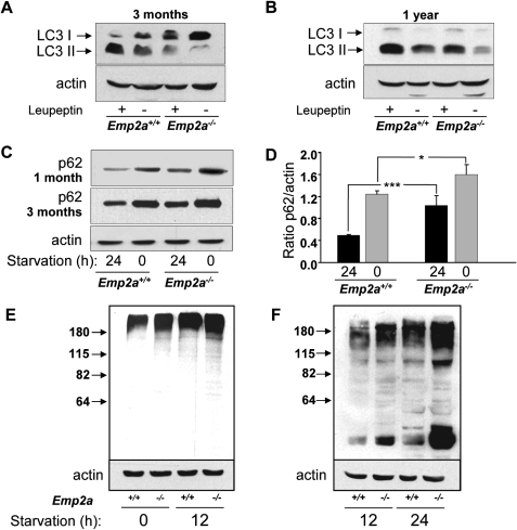
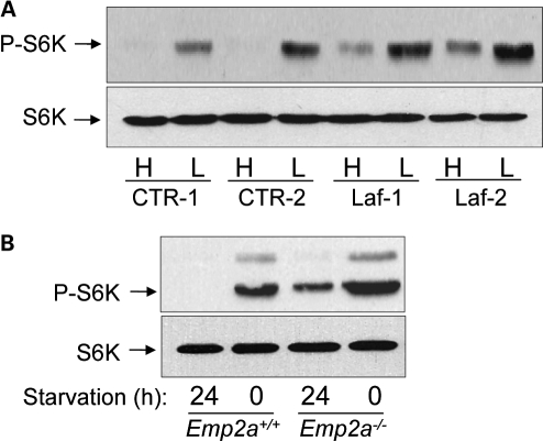
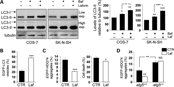
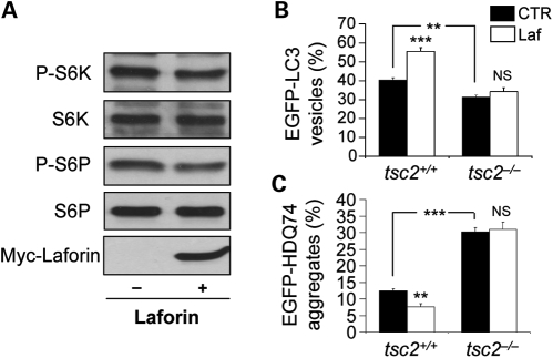
Lafora disease (LD) is an autosomal recessive, progressive myoclonus epilepsy, which is characterized by the accumulation of polyglucosan inclusion bodies, called Lafora bodies, in the cytoplasm of cells in the central nervous system and in many other organs. However, it is unclear at the moment whether Lafora bodies are the cause of the disease, or whether they are secondary consequences of a primary metabolic alteration. Here we describe that the major genetic lesion that causes LD, loss-of-function of the protein laforin, impairs autophagy. This phenomenon is confirmed in cell lines from human patients, mouse embryonic fibroblasts from laforin knockout mice and in tissues from such mice. Conversely, laforin expression stimulates autophagy. Laforin regulates autophagy via the mammalian target of rapamycin kinase-dependent pathway. The changes in autophagy mediated by laforin regulate the accumulation of diverse autophagy substrates and would be predicted to impact on the Lafora body accumulation and the cell stress seen in this disease that may eventually contribute to cell death.


Figures

Figure 1.
Figure 1.
Loss of laforin decreases formation of autophagosomes. (A, B, D, E) Representative immunoblots, using antibodies which recognize LC3 or, as a loading control, actin, with extracts (75 µg protein) of human fibroblasts from a control individual (CTR-1) and from two different LD patients (Laf-1 and Laf-2) (A and B), or of MEFs from control (Epm2a+/+) and laforin-deficient (Epm2a−/−) mice (D and E), incubated for 2 h without (A and D) and with (B and E) bafilomycin A1 (400 nM) under conditions of high (H, Krebs-Henseleit medium) and low (L, full medium) proteolysis. The positions of LC3-I and LC3-II bands are indicated on the left. (C and F) The LC3-II bands from three independent experiments similar to those shown in (B) and (E), respectively, were quantified by densitometry and normalized to the corresponding actin bands. Data are expressed in percent relative to control cells.
Figure 2.
Figure 2.
Loss of laforin decreases the degradation of long-lived proteins by macroautophagy and increases accumulation of polyubiquitinated proteins. (A and B) Intracellular protein degradation (total) and the amount of protein degraded by macroautophagy (MA) was calculated as described in Materials and Methods in control (CTR, black histograms) and laforin-deficient (Laf-, white histograms) human fibroblasts (A) and in MEFs from control (Epm2a+/+, black histograms) and laforin-deficient (Epm2a−/−, white histograms) mice (B). Each value represents the mean from three different experiments with duplicated samples, using in each case two different control cell lines and two different laforin-deficient cells. (C and D) Extracts (75 µg protein) of fibroblasts from a representative control individual (CTR-1) and from two different LD patients (Laf-1 and Laf-2), incubated for 2 h, without (C) or with (D) the proteasomal inhibitor MG-132 (20 µM) under conditions which produce high (H, Krebs–Henseleit medium) and low (L, full medium) proteolysis, were analysed on western blots using antibodies that recognize ubiquitinated proteins and actin, which serves as a loading control. The positions of molecular-mass markers and their size in kDa are indicated on the left. Upper gels: low exposure (exp.), lower gels: high exposure.
Figure 3.
Figure 3.
Loss of laforin slows macroautophagy in vivo. (A and B) Representative immunoblots using anti-LC3 or anti-actin of mouse liver lysates (100 µg protein) from control (Epm2a+/+) and laforin-deficient (Epm2a−/−) mice, 3 months (A) and 1 year (B) old, starved for 24 h and injected (+) or not (−) with 2 mg/100 g weight leupeptin. The positions of LC3-I and LC3-II bands are indicated. (C) Mouse liver lysates (50 µg protein) of 1-month and 3-month-old mice, starved for 24 h (high proteolysis) or fed (0, low proteolysis), were immunoblotted with p62 and actin antibodies. (D) The bands from four independent experiments (two from each 1-month and 3-month-old mice) similar to those shown in (C) were assessed by densitometry and normalized to the corresponding actin bands. (E and F) Control (Epm2a+/+) and laforin-deficient (Epm2a−/−) mice, 1 (E) and 3 (F) months old, were fed ad libitum (0) or fasted (starvation) for 12 or 24 h. Mouse liver lysates (100 µg protein) were analysed on western blots using antibodies that recognize ubiquitinated proteins and actin, which serves as a loading control. The positions of molecular-mass markers and their size in kDa are indicated on the left.
Figure 4.
Figure 4.
mTOR signalling pathway is upregulated in laforin-deficient cells. (A) Human fibroblasts from two control individuals (CTR-1 and CTR-2) and from two different LD patients (Laf-1 and Laf-2) were incubated for 2 h under conditions of high (H, Krebs–Henseleit medium) and low (L, full medium) proteolysis in the cells. Then, extracts (75 µg) were prepared to determine the activation state of the down-stream target of mTOR p70S6 kinase (S6K) by immunoblot analysis, using antibodies which recognize phosphorylated Thr389 in p70S6 kinase (P-S6K) and total S6K. (B) mTOR activation was also analysed by the same procedures in liver extracts from 3-month-old control (Epm2a+/+) and laforin-deficient (Epm2a−/−) mice, starved for 24 h (high proteolysis conditions) or fed (0, low proteolysis conditions).
Figure 5.
Figure 5.
Wild-type laforin induces autophagy and facilitates the clearance of autophagy substrates. (A) COS-7 or SK-N-SH cells, transfected with 2 µg pcDNA3.1 (empty vector) or Myc-Laforin for 4 h, were treated with or without 400 nM bafilomycin A1 in the last 4 h of the 24 h post-transfection period. Overexpression of wild-type laforin increased autophagosome synthesis, as analysed by immunoblotting with anti-LC3 antibody (upper gels: low exposure (exp.), lower gels: high exposure) and densitometric analysis of LC3-II levels relative to tubulin. (B) COS-7 cells, transfected with 0.5 µg EGFP-LC3 and either 1.5 µg pcDNA3.1 (empty vector) or Myc-Laforin for 4 h, were fixed and analysed for EGFP-LC3 vesicles at 24 h post-transfection. Overexpression of wild-type laforin increased the proportion of transfected (EGFP-positive) cells with EGFP-LC3 vesicles. (C) COS-7 cells, transfected with 0.5 µg EGFP-HDQ74 and either 1.5 µg pcDNA3.1 (empty vector) or Myc-Laforin for 4 h, were fixed and analysed for EGFP-HDQ74 aggregates and cell death at 48 h post-transfection. Overexpression of wild-type laforin reduced the percentages of tranfected (EGFP-positive) cells with mutant huntingtin aggregates and cell death, assessed by apoptotic nuclear morphology (see Materials and Methods). (D) atg5+/+ and atg5−/− MEFs, transfected with 0.5 µg EGFP-HDQ74 and either 1.5 µg pcDNA3.1 (empty vector) or Myc-Laforin for 4 h, were fixed and analysed for EGFP-HDQ74 aggregates at 48 h post-transfection. Overexpression of wild-type laforin reduced mutant huntingtin aggregates in atg5+/+ MEFs, but not in atg5−/− MEFs. atg5−/− MEFs had increased aggregates compared with atg5+/+ MEFs.
Figure 6.
Figure 6.
Wild-type laforin reduces mTOR activity to regulate autophagy. (A) COS-7 cells, transfected with 2 µg pcDNA3.1 (empty vector) or Myc-Laforin for 4 h, were analysed for mTOR activity at 24 h post-transfection by immunoblotting with anti-phospho-S6 kinase (P-S6K, Thr389) and anti-phospho-S6 ribosomal protein (P-S6P, Ser235/236) antibodies. Overexpression of wild-type laforin (detected with anti-myc antibody) reduced phosphorylation of S6K and S6P relative to the total proteins. (B) tsc2+/+ and tsc2−/− MEFs, transfected with 0.5 µg EGFP-LC3 and either 1.5 µg pcDNA3.1 (empty vector) or Myc-Laforin for 4 h, were fixed and analysed for EGFP-LC3 vesicles at 24 h post-transfection. Overexpression of wild-type laforin increased the proportion of cells with EGFP-LC3 vesicles in tsc2+/+ MEFs, but not in tsc2−/− MEFs. tsc2−/− MEFs had a lower proportion of cells with EGFP-LC3 vesicles compared with tsc2+/+ MEFs. (C) tsc2+/+ and tsc2−/− MEFs, transfected with 0.5 µg EGFP-HDQ74 and either 1.5 µg pcDNA3.1 (empty vector) or Myc-Laforin for 4 h, were fixed and analysed for EGFP-HDQ74 aggregates at 48 h post-transfection. Overexpression of wild-type laforin reduced mutant huntingtin aggregates in tsc2+/+ MEFs, but not in tsc2−/− MEFs. tsc2−/− MEFs had increased aggregates compared with tsc2+/+ MEFs.
Figure 7.
Figure 7.
Schematic representation of regulation of autophagy by laforin and the disease consequences. Wild-type laforin induces autophagy by inhibiting mTOR in a TSC2-dependent manner. Loss of laforin in LD activates mTOR and inhibits autophagy. An impairment of autophagy in LD may contribute to the disease pathogenesis (indicated in dashed line).

References

    1. Lafora G.R., Glueck B. Beitrag zur Histopathologie der myoklonischen Epilepsie. Z. Gesammte Neurol. Psychiatr. 1911;6:1–14. doi:10.1007/BF02863929. - DOI
    1. Van Hoof F., Hageman-Bal M. Progressive familial myoclonic epilepsy with Lafora bodies. Electron microscopic and histochemical study of a cerebral biopsy. Acta Neuropathol. 1967;7:315–336. - PubMed
    1. Yokoi S., Nakayama H., Negishi T. Biochemical studies on tissues from a patient with Lafora disease. Clin. Chim. Acta. 1975;62:415–423. - PubMed
    1. Ganesh S., Delgado-Escueta A.V., Suzuki T., Francheschetti S., Riggio C., Avanzini G., Rabinowicz A., Bohlega S., Bailey J., Alonso M.E., et al. Genotype-phenotype correlations for EPM2A mutations in Lafora's progressive myoclonus epilepsy: exon 1 mutations associate with an early-onset cognitive deficit subphenotype. Hum. Mol. Genet. 2002;11:1263–1271. doi:10.1093/hmg/11.11.1263. - DOI - PubMed
    1. Sinha S., Satishchandra P., Gayathri N., Yasha T.C., Shankar S.K. Progressive myoclonic epilepsy: a clinical, electrophysiological and pathological study from South India. J. Neurol. Sci. 2007;252:16–23. doi:10.1016/j.jns.2006.09.021. - DOI - PubMed

Publication types

MeSH terms

Substances