. 2010 Nov;192(21):5755-66.
doi: 10.1128/JB.00135-10. Epub 2010 May 7.

The hyperthermophilic euryarchaeon Archaeoglobus fulgidus repairs uracil by single-nucleotide replacement

Affiliations

The hyperthermophilic euryarchaeon Archaeoglobus fulgidus repairs uracil by single-nucleotide replacement

Ingeborg Knævelsrud et al. J Bacteriol. 2010 Nov.

Abstract

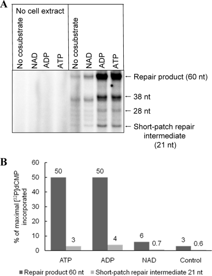
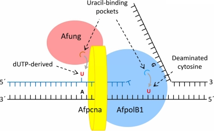
Hydrolytic deamination of cytosine to uracil in cellular DNA is a major source of C-to-T transition mutations if uracil is not repaired by the DNA base excision repair (BER) pathway. Since deamination increases rapidly with temperature, hyperthermophiles, in particular, are expected to succumb to such damage. There has been only one report of crenarchaeotic BER showing strong similarities to that in most eukaryotes and bacteria for hyperthermophilic Archaea. Here we report a different type of BER performed by extract prepared from cells of the euryarchaeon Archaeoglobus fulgidus. Although immunodepletion showed that the monofunctional family 4 type of uracil-DNA glycosylase (UDG) is the principal and probably only UDG in this organism, a β-elimination mechanism rather than a hydrolytic mechanism is employed for incision of the abasic site following uracil removal. The resulting 3' remnant is removed by efficient 3'-phosphodiesterase activity followed by single-nucleotide insertion and ligation. The finding that repair product formation is stimulated similarly by ATP and ADP in vitro raises the question of whether ADP is more important in vivo because of its higher heat stability.


Figures

FIG. 1.
FIG. 1.
Dependence on protein for excision of uracil from DNA by A. fulgidus cell extract (A) and rAfung (B). Different amounts of cell extract (A) or rAfung (B) were incubated with [3H]uracil-containing DNA (2,400 dpm; 2.2 pmol DNA uracil residues) in reaction buffer at 80°C for 10 min. Each value is the average of two independent measurements.
FIG. 2.
FIG. 2.
Immunodepletion of the uracil-releasing activity present in A. fulgidus cell extract. Protein extract (240 μg) (in panel A also purified enzyme [0.88 μg]) was treated with different volumes of rabbit antiserum plus reaction buffer in a 500-μl (total volume) mixture, which was followed by centrifugation as described in Materials and Methods. (A) Supernatant following the immunodepletion procedure (25 μl) was incubated for 10 min with [3H]uracil-containing DNA (2,000 dpm; 2 pmol DNA uracil residues) in reaction buffer at 80°C. Symbols: •, cell extract treated with anti-rAfungH; ○, cell extract treated with control serum; □, rAfungH′ (rAfungH following enzymatic removal of the His tag) treated with anti-rAfungH. Each value is the mean of three independent measurements. The lower graph is an expansion of the 0-to-1 μl part of the upper graph. (B) Supernatant following immunodepletion (5 μl) was incubated with substrate 1 (32P-labeled 5′-TAGACATTGCCCTCGAGGTAUCATGGATCCGATTTCGACCTCAAACCTAGACGAATTCCG-3′ plus complementary strand; 4 fmol) at 60°C for 10 min in reaction buffer (20 μl). As a result of uracil excision and base-catalyzed phosphodiester bond cleavage (generating a mixture of β- and δ-elimination products; see Fig. 7A), each 32P-end-labeled oligonucleotide (60 nt) was converted into one 32P-labeled 20-nt product and one 40-nt unlabeled product. The strand opposite the damage-containing strand is indicated by a dashed line; the sizes of the 32P-labeled repair intermediates and products are indicated by solid lines. CS, control serum.
FIG. 3.
FIG. 3.
Western blot analysis of the Afung protein present in A. fulgidus cell extract. Following SDS-PAGE of A. fulgidus cell extract and purified rAfung and rAfungH (the results of one such experiment performed with a 12% [wt/vol] polyacrylamide gel stained with Coomassie blue are shown in panel A), Western blot analysis (B) using antiserum raised against rAfungH (left panel, diluted 1/4,000; right panel, diluted 1/1,000) was performed with purified rAfung and rAfungH and cell extract as described in Materials and Methods.
FIG. 4.
FIG. 4.
Lineweaver-Burk plots for excision of uracil from DNA by wtAfung (A) and rAfung (B). Protein extract (14 μg containing 0.33 pmol wtAfung) or rAfung (6.2 pmol) was incubated with different amounts of [3H]uracil-containing DNA (7.55 to 75.5 pmol DNA uracil residues) in reaction buffer at pH 7.5 for 10 min at 80°C. Each value is the average of two to six independent measurements.
FIG. 5.
FIG. 5.
Sequence alignment of Afung, Tmung of T. maritima, and Ttung of T. thermophilus (A), Afung protein structure (B), and structure of the Afung substrate-binding site with a uracil base inserted (C). (A) Secondary structure elements of Tmung (also designated TmUDG [Table 2]) and Ttung (also designated TtUDGa [Table 2]) are indicated above and below the alignment, respectively. α-Helices are indicated by spirals, and β-strands are indicated by arrows. Identical residues in all of the sequences and identical residues in two of the sequences are indicated by white letters and red letters, respectively. Residues indicated by a blue asterisk are the cysteines that coordinate the iron-sulfur cluster present in the family 4 UDGs. The residue numbering is consistent with Afung numbering. The sequences were aligned using ClustalW as described in Materials and Methods. (B) Cartoon showing the overall homology model of Afung. The secondary structure elements are shown; α-helices are indicated by blue spirals, β-strands are indicated by red arrows, and the loop structure is indicated by green tubes. The iron-sulfur cluster was modeled based on the structure of Ttung, and black and yellow spheres represent iron and sulfur, respectively. The coordinating cysteine residues are indicated by sticks. Uracil in the substrate-binding pocket, based on the structure of Ttung, is also indicated by a stick representation. The cartoon was drawn using PyMOL (10). (C) Close-up of the substrate-binding pocket of Afung and the putative interactions with uracil, showing uracil with orange carbon atoms and protein residues with green carbon atoms. Other atoms are also shown (blue, nitrogen; red, oxygen). Hydrogen bonds involving uracil are indicated by black dashed lines. Repulsion between side chain atoms of Glu48 and a substituent in the C-5 position in uracil is indicated by a dashed red line. The cartoon was drawn using PyMOL (10).
FIG. 6.
FIG. 6.
Diagram of the steps of the BER pathway of A. fulgidus for repair of uracil in DNA. The residues removed and the results of replacement are indicated by red and blue, respectively; proteins with verified biochemical activity (e.g., Afung and Afogg; however, whether Afxth contains 3′-phosphodiesterase activity and whether AfpolB1 is able to fill in one-nucleotide gaps have not been verified [6, 52]) are indicated by black type; putative proteins based on genomic sequences (ORFs) are indicated by white type. The genes encoding the proteins are as follows (Swiss-Prot/TrEMBL accession numbers are indicated in parentheses): Afogg, AF0371 (O29876); Afnth, AF1692 (O28581); Afxth, AF0580 (O29675); AfpolB1, AF0497 (O29753); AfpolB2, AF0693m; Aflig1, AF0623 (O29632); and Aflig2, AF1725 (O28549). dR, deoxyribose residue; dR′, modified deoxyribose residue (i.e., α,β-unsaturated aldehyde); dRP, deoxyribose phosphate; P, phosphate group. A solid arrow indicates a step confirmed experimentally; a dashed arrow indicates a step not demonstrated experimentally yet.
FIG. 7.
FIG. 7.
Experimental verification of the different steps of the BER pathway of A. fulgidus for repair of uracil in DNA. The 3′ (A) and 5′ (B) incision products and the short-patch repair intermediate and product (C) were determined following excision of uracil from DNA by A. fulgidus cell extract. (A and B) Protein extract (11.4 μg) was incubated with 0.177 pmol substrate 1 (A) (see Fig. 2B) or 2.475 fmol substrate 2 (sequence identical to substrate 1 sequence except for an additional dCMP at the 3′ end as the 32P label; complementary strand identical to that of substrate 1) (B) at 60°C for 10 min in 45 mM HEPES-KOH (pH 7.8), 0.4 mM EDTA, 1 mM DTT, 2% (vol/vol) glycerol, 70 mM KCl with or without 5 mM MgCl2 (total volume, 10 μl) with E. coli enzymes (1 U of Ung; 1 μg of Nfo, Nth, or Fpg) at 37°C for 10 min in buffer without MgCl2. 5′-dRP, 5′-deoxyribose phosphate; 3′-OH, AP endonuclease product; 5′-P, 5′-phosphate; β, β-elimination product (α,β-unsaturated aldehyde); δ, δ-elimination product; *, alkaline phosphatase treatment. (C) Results of a typical experiment showing conversion of repair intermediates to repair product as a function of time. Substrate 3 (0.5 pmol), which is an unlabeled version of substrate 1, was incubated with A. fulgidus protein extract (14 μg) at 60°C in 45 mM HEPES-KOH (pH 7.8), 0.4 mM EDTA, 1 mM DTT, 2% (vol/vol) glycerol, 70 mM KCl, 5 mM MgCl2, 1 μg/μl BSA, 13 μM dATP, 13 μM dTTP, 13 μM dGTP, [α-32P]dCTP in a 15-μl (final volume) mixture. The lower panel shows the relative concentrations of the short-patch repair intermediate and repair product as a function of time based on the average values of two independent measurements.
FIG. 8.
FIG. 8.
Effect of nucleotides on BER of uracil in DNA by A. fulgidus cell extract. (A) Results of a typical experiment comparing ATP, ADP, and NAD as stimulatory agents for conversion of a repair intermediate to a repair product following 60 min of incubation. (B) Average concentrations of repair intermediate and repair product as determined in three independent experiments, as shown in panel A. Substrate 3 (0.5 pmol) was incubated with A. fulgidus protein extract (17 μg) at 60°C in 45 mM HEPES-KOH (pH 7.8), 0.4 mM EDTA, 1 mM DTT, 70 mM KCl, 5 mM MgCl2, 2% (vol/vol) glycerol, 1 μg/μl BSA, 13 μM dATP, 13 μM dTTP, 13 μM dGTP, [α-32P]dCTP in a 15-μl (final volume) mixture.
FIG. 9.
FIG. 9.
Model for Afung-initiated repair of misincorporated uracil and detection of deaminated cytosine in DNA during replication. Afung contains a PCNA-binding motif (69), indicating that there is PCNA-dependent postreplicative removal of uracil, where Afpcna (AF0335 [accession no. O29912]) is a homotrimer. Interactions of AfpolB1 with Afpcna have been demonstrated experimentally (42, 53, 54). Detection and accommodation of uracil by AfpolB1 probably interrupt chain elongation, which may promote reassembly of template strands and transfer of Afung to the lesion site initiating BER, as shown in Fig. 6. See text for further details.

Similar articles

Cited by

References

    1. Akbari, M., M. Otterlei, J. Peña-Diaz, P. A. Aas, B. Kavli, N. B. Liabakk, L. Hagen, K. Imai, A. Durandy, G. Slupphaug, and H. E. Krokan. 2004. Repair of U/G and U/A in DNA by UNG2-associated repair complexes takes place predominantly by short-patch repair both in proliferating and growth-arrested cells. Nucleic Acids Res. 32:5486-5498. - PMC - PubMed
    1. Alseth, I., H. Korvald, F. Osman, E. Seeberg, and M. Bjørås. 2004. A general role of the DNA glycosylase Nth1 in the abasic sites cleavage step of base excision repair in Schizosaccharomyces pombe. Nucleic Acids Res. 32:5119-5125. - PMC - PubMed
    1. Andersen, S., T. Heine, R. Sneve, I. König, H. E. Krokan, B. Epe, and H. Nilsen. 2005. Incorporation of dUMP into DNA is a major source of spontaneous DNA damage, while excision of uracil is not required for cytotoxicity of fluoropyrimidines in mouse embryonic fibroblasts. Carcinogenesis 26:547-555. - PubMed
    1. Birkeland, N.-K., H. Ånensen, I. Knævelsrud, W. Kristoffersen, M. Bjørås, F. T. Robb, A. Klungland, and S. Bjelland. 2002. Methylpurine DNA glycosylase of the hyperthermophilic archaeon Archaeoglobus fulgidus. Biochemistry 41:12697-12705. - PubMed
    1. Bradford, M. M. 1976. A rapid and sensitive method for the quantitation of microgram quantities of protein utilizing the principle of protein-dye binding. Anal. Biochem. 72:248-254. - PubMed

Publication types

MeSH terms

LinkOut - more resources