Pregnancy-related changes in connections from the cervix to forebrain and hypothalamus in mice
- PMID: 20453158
- PMCID: PMC4242598
- DOI: 10.1530/REP-10-0002
Pregnancy-related changes in connections from the cervix to forebrain and hypothalamus in mice
Abstract
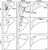
The transneuronal tracer pseudorabies virus was used to test the hypothesis that connections from the cervix to the forebrain and hypothalamus are maintained with pregnancy. The virus was injected into the cervix of nonpregnant or pregnant mice, and, after 5 days, virus-labeled cells and fibers were found in specific forebrain regions and, most prominently, in portions of the hypothalamic paraventricular nucleus. With pregnancy, fewer neurons and fibers were evident in most brain regions compared to that in nonpregnant mice. In particular, little or no virus was found in the medial and ventral parvocellular subdivisions, anteroventral periventricular nucleus, or motor cortex in pregnant mice. By contrast, labeling of virus was sustained in the dorsal hypothalamus and suprachiasmatic nucleus in all groups. Based upon image analysis of digitized photomicrographs, the area with label in the rostral and medial parvocellular paraventricular nucleus and magnocellular subdivisions was significantly reduced in mice whose cervix was injected with virus during pregnancy than in nonpregnant mice. The findings indicate that connections from the cervix to brain regions that are involved in sensory input and integrative autonomic functions are reduced during pregnancy. The findings raise the possibility that remaining pathways from the cervix to the forebrain and hypothalamus may be important for control of pituitary neuroendocrine secretion, as well as for effector functions in the cervix as pregnancy nears term.
Conflict of interest statement
The authors declare that there is no conflict of interest that could be perceived as prejudicing the impartiality of the research reported.
Figures


References
-
- Akaishi T, Robbins A, Sakuma Y, Sato Y. Neural inputs from the uterus to the paraventricular magnocellular neurons in the rat. Neuroscience Letters. 1988;84:57–62. - PubMed
-
- Apostolakis EM, Rice KE, Longo LD, Seron-ferre M, Yellon SM. Time of day of birth and absence of endocrine and uterine contractile activity rhythms in sheep. American Journal of Physiology. 1993;264:E534–E540. - PubMed
-
- Armstrong WE. Hypothalamic supraoptic and paraventricular nuclei. In: Paxinos G, editor. The Rat Nervous System. San Diego, CA: Academic Press; 1995. pp. 377–390.
-
- Aston-Jones G, Card JP. Use of pseudorabies virus to delineate multisynaptic circuits in brain: opportunities and limitations. Journal of Neuroscience Methods. 2000;103:51–61. - PubMed
-
- Bittencourt JC, Benoit R, Sawchenko PE. Distribution and origins of substance P-immunoreactive projections to the paraventricular and supraoptic nuclei: partial overlap with ascending catecholaminergic projections. Journal of Chemical Neuroanatomy. 1991;4:63–78. - PubMed
Publication types
MeSH terms
Grants and funding
LinkOut - more resources
Full Text Sources
Medical

