. 2010 Aug;299(2):L150-9.
doi: 10.1152/ajplung.00341.2009. Epub 2010 May 7.

Nitric oxide synthase 3 contributes to ventilator-induced lung injury

Affiliations

Nitric oxide synthase 3 contributes to ventilator-induced lung injury

Katerina Vaporidi et al. Am J Physiol Lung Cell Mol Physiol. 2010 Aug.

Abstract

Nitric oxide synthase (NOS) depletion or inhibition reduces ventilator-induced lung injury (VILI), but the responsible mechanisms remain incompletely defined. The aim of this study was to elucidate the role of endothelial NOS, NOS3, in the pathogenesis of VILI in an in vivo mouse model. Wild-type and NOS3-deficient mice were ventilated with high-tidal volume (HV(T); 40 ml/kg) for 4 h, with and without adding NO to the inhaled gas. Additional wild-type mice were pretreated with tetrahydrobiopterin and ascorbic acid, agents that can prevent NOS-generated superoxide production. Arterial blood gas tensions, histology, and lung mechanics were evaluated after 4 h of HV(T) ventilation. The concentration of protein, IgM, cytokines, malondialdehyde, and 8-isoprostane were measured in bronchoalveolar lavage fluid (BALF). Myeloperoxidase activity, total and oxidized glutathione levels, and NOS-derived superoxide production were measured in lung tissue homogenates. HV(T) ventilation induced VILI in wild-type mice, as reflected by decreased lung compliance, increased concentrations of protein and cytokines in BALF, and oxidative stress. All indices of VILI were ameliorated in NOS3-deficient mice. Augmenting pulmonary NO levels by breathing NO during mechanical ventilation did not increase lung injury in NOS3-deficient mice. HV(T) ventilation increased NOS-inhibitable superoxide production in lung extracts from wild-type mice but not in those from NOS3-deficient mice. Administration of tetrahydrobiopterin and ascorbic acid ameliorated VILI in wild-type mice. Our results indicate that NOS3 contributes to ventilator-induced lung injury via increased production of superoxide.


Figures

Fig. 1.
Fig. 1.
A: peak inspiratory pressure (PIP) during high-tidal volume (HVT) ventilation (VT 40 ml/kg) in wild-type (WT) mice (gray) and NOS3−/− mice (white), without (circles) or with 5 ppm (diamonds) or 50 ppm (triangles) inhaled NO, and BH4/ascorbic acid-treated WT mice (squares). During the first 60 min of mechanical ventilation at tidal volume (VT) 8 ml/kg, PIP was 8–9 cmH2O in all mice. PIP increased during HVT ventilation in WT mice not receiving inhaled NO and in WT mice receiving 50 ppm inhaled NO (*P < 0.05 vs. all other groups). For clarity, PIP only during HVT ventilation is shown. B: inspiratory pressure-volume curve of the respiratory system in WT mice (gray) and NOS3−/− mice (white), in control mice (plus symbol) and in mice after HVT ventilation without (circles), or with 5 ppm (diamonds) or 50 ppm (triangles) inhaled NO, and WT mice treated with BH4 and ascorbic acid (squares). HVT ventilation resulted in deterioration of lung mechanics in all ventilated mice (P < 0.001 WT-HVT vs. WT control, P < 0.01 NOS3−/−-HVT vs. NOS3−/− control). Lung compliance decreased to a greater extent in WT mice than in NOS3−/− mice (P < 0.01 NOS3−/−-HVT vs. WT-HVT). Inhaled NO at 5 or 50 ppm did not affect lung mechanics in NOS3−/− mice subjected to HVT. In WT mice, breathing 5 ppm NO improved lung mechanics (P < 0.01 WT-HVT+iNO5ppm vs. WT-HVT), whereas breathing 50 ppm NO did not. Treatment of WT mice exposed to HVT with BH4 and ascorbic acid improved lung mechanics (P < 0.01 WT-HVT+BH4/asc vs. WT-HVT). For clarity, only mean values are shown. N = 5–10 per group.
Fig. 2.
Fig. 2.
Protein concentration in BALF from control WT and NOS3−/− mice, and WT and NOS3−/− mice ventilated with HVT (40 ml/kg) for 4 h, without or with inhaled NO 5 ppm (iNO5ppm) or 50 ppm (iNO50ppm). BALF protein levels were greater in WT and NOS3−/− mice subjected to HVT ventilation than in the corresponding controls (*P < 0.001 and #P < 0.05, respectively). BALF protein concentrations after HVT ventilation were less in NOS3−/− mice and in WT mice breathing 5 ppm NO than in WT mice (†P < 0.001). N = 7–10 per group. Boxes represent 25th-75th percentile; line and square represent median and mean, respectively; whisker represents 5th-95th percentile, and × represents 1st-99th percentile.
Fig. 3.
Fig. 3.
IgM concentrations in BALF from control WT and NOS3−/− mice and from WT and NOS3−/− mice ventilated with HVT (40 ml/kg) for 4 h. BALF IgM levels were greater in WT and NOS3−/− mice subjected to HVT ventilation than in the corresponding controls (*P < 0.001 and #P < 0.05, respectively). BALF IgM concentration after HVT ventilation was less in NOS3−/− mice than in WT mice (†P < 0.001). N = 7–10 per group. Data are presented in box plots as in Fig. 2.
Fig. 4.
Fig. 4.
IL-6 concentrations in BALF from control WT and NOS3−/− mice and WT and NOS3−/− mice ventilated with HVT (40 ml/kg) for 4 h, without or with inhaled NO 5 ppm (iNO5ppm) or 50 ppm (iNO50ppm). BALF IL-6 concentrations were greater in WT and NOS3−/− mice subjected to HVT ventilation than in the corresponding controls (*P < 0.001). BALF IL-6 concentrations after HVT ventilation were less in NOS3−/− mice and in WT mice breathing 5 ppm NO than in WT mice not receiving NO (#P < 0.001). BALF IL-6 concentrations after HVT ventilation were greater in WT mice breathing 50 ppm NO than in WT mice not receiving NO (†P < 0.01). N = 3–4 per group as controls and 6–10 per group for HVT ventilation. Data are presented in box plots as in Fig. 2.
Fig. 5.
Fig. 5.
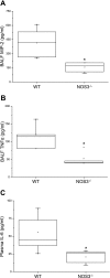
MIP-2 (A) and TNFα (B) concentrations in BALF, and IL-6 concentrations in plasma (C) from WT and NOS3−/− mice ventilated with HVT (40 ml/kg) for 4 h. In control mice of both genotypes, MIP-2 and TNFα were not detected in BALF, and IL-6 was not detected in plasma. After HVT ventilation, BALF MIP-2 and TNFα concentrations were increased in both genotypes, but less in NOS3−/− mice than in WT mice (*P < 0.01). Plasma IL-6 concentrations after HVT ventilation were less in NOS3−/− mice than in WT mice (*P < 0.001). N = 3–4 per group as controls, and 6–10 per group for HVT ventilation. Data are presented in box plots as in Fig. 2.
Fig. 6.
Fig. 6.
Lung myeloperoxidase (MPO) activity from control WT and NOS3−/− mice and WT and NOS3−/− mice ventilated with HVT (40 ml/kg) for 4 h, with or without 5 ppm inhaled NO (iNO5ppm). Lung MPO activity was greater in WT and NOS3−/− mice subjected to HVT ventilation than in the corresponding controls (*P < 0.001 and #P < 0.01, respectively). Lung MPO activity after HVT ventilation was less in NOS3−/− mice and in WT mice breathing 5 ppm NO than in WT mice not receiving NO (†P < 0.01 and ‡P < 0.001, respectively). Data are expressed as MPO activity units per milligram of tissue; n = 3–4 per group for control and 6–9 per group for HVT ventilation. Data are presented in box plots as in Fig. 2.
Fig. 7.
Fig. 7.
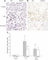
Histology and immunohistochemistry for neutrophils in lung sections from control WT and NOS3−/− mice and from WT and NOS3−/− mice subjected to 4 h of HVT ventilation (40 ml/kg) with or without 5 ppm (iNO5ppm) inhaled NO. A: representative photographs of lung sections stained with hematoxylin and eosin and presented in ×40 original magnification. Control WT (1) and NOS3−/− (2) mice had preserved lung parenchymal architecture, and there were no differences observed between genotypes. HVT ventilation resulted in increased cellular infiltration and alveolar wall thickening in WT mice (3) that was less marked in NOS3−/− mice (4) and in WT mice breathing 5 ppm NO (5). Breathing 5 ppm NO did not alter the histological appearance of lungs from NOS3−/− mice exposed to HVT ventilation (6). B: representative photographs of lung sections reacted with anti-mouse neutrophil monoclonal antibody are presented in ×40 original magnification. Few neutrophils (arrows) were observed in lungs from control WT (7) and NOS3−/− (8) mice. HVT ventilation induced neutrophil infiltration to a greater degree in WT (9) than in NOS3−/− (10) mice. Breathing 5 ppm NO reduced neutrophil accumulation in WT mice subjected to HVT ventilation (11). Neutrophil infiltration after HVT ventilation was similar in NOS3−/− with (12) and without inhaled NO. White arrow shows neutrophils inside small vessels. C: quantification of neutrophil immunostaining in lungs from WT and NOS3−/− mice. Neutrophil infiltration was greater in WT and NOS3−/− mice subjected to HVT ventilation than in the corresponding controls (*P < 0.001); neutrophil infiltration after HVT ventilation was less in NOS3−/− mice and in WT mice breathing 5 ppm NO than in WT mice not receiving NO (#P < 0.01 for both). Data represent means ± SD of number of neutrophils per high-power magnification field counted in at least 50 fields (×40) for each group.
Fig. 8.
Fig. 8.
BALF concentrations of NO metabolites (nitrite/nitrate, NOx) from control WT and NOS3−/− mice and WT and NOS3−/− mice ventilated with HVT (40 ml/kg) for 4 h. BALF NOx levels were greater in WT mice subjected to HVT ventilation than in WT controls (*P < 0.001). BALF NOx levels after HVT ventilation were less in NOS3−/− mice than in WT mice (#P < 0.01). N = 5–8 per group. Data are presented in box plots as in Fig. 2.
Fig. 9.
Fig. 9.
A: BALF concentrations of malondialdehyde (MDA) from control WT and NOS3−/− mice and WT and NOS3−/− mice ventilated with HVT (40 ml/kg) for 4 h. BALF MDA levels were greater in WT mice subjected to HVT ventilation than in WT controls (*P < 0.01), but were unchanged in NOS3−/− mice. BALF MDA levels after HVT ventilation were less in NOS3−/− mice than in WT mice (#P < 0.01). Data are presented in box plots as in Fig. 2; n = 6–8 per group. B: BALF concentrations of 8-isoprostane from control WT and NOS3−/− mice and WT and NOS3−/− mice ventilated with HVT (40 ml/kg) for 4 h. BALF 8-isoprostane levels were greater in WT and NOS3−/− mice subjected to HVT ventilation than in corresponding controls (*P < 0.001 and #P < 0.01, respectively). BALF MDA levels after HVT ventilation were less in NOS3−/− than WT mice (†P < 0.01). Data are presented in box plots as in Fig. 2; n = 5 per group. C: the ratio of total to oxidized glutathione levels (GSH/GSSG) in lung tissues from control WT and NOS3−/− mice, and WT and NOS3−/− mice ventilated with HVT (40 ml/kg) for 4 h. GSH/GSSG was less in WT mice subjected to HVT ventilation than in WT controls (*P < 0.05). HVT ventilation did not reduce GSH/GSSG in NOS3−/− mice, and GSH/GSSG was greater in NOS3−/− mice subjected to HVT ventilation than in similarly ventilated WT mice (*P < 0.05). Data are presented in box plots as in Fig. 2; n = 5 per group. D: NOS-derived superoxide production in lung homogenates from control WT and NOS3−/− mice and WT and NOS3−/− mice ventilated with HVT (40 ml/kg) for 4 h. NOS-derived superoxide production was estimated from the difference in lucigenin-enhanced chemiluminescence (LEC) measured in the absence and presence of l-NAME (1 mM). l-NAME-inhibitable superoxide production was greater in WT mice subjected to HVT ventilation than in WT control mice and in similarly ventilated NOS3−/− mice (*P < 0.01 for both). Data are expressed as relative light units (RLU) per second per milligram of protein, n = 5 per group. Data are presented in box plots as in Fig. 2.

Comment in

References

    1. Abdulnour RE, Peng X, Finigan JH, Han EJ, Hasan EJ, Birukov KG, Reddy SP, Watkins JE, 3rd, Kayyali US, Garcia JG, Tuder RM, Hassoun PM. Mechanical stress activates xanthine oxidoreductase through MAP kinase-dependent pathways. Am J Physiol Lung Cell Mol Physiol 291: L345–L353, 2006 - PubMed
    1. Altemeier WA, Matute-Bello G, Gharib SA, Glenny RW, Martin TR, Liles WC. Modulation of lipopolysaccharide-induced gene transcription and promotion of lung injury by mechanical ventilation. J Immunol 175: 3369–3376, 2005 - PubMed
    1. Antoniades C, Shirodaria C, Warrick N, Cai S, de Bono J, Lee J, Leeson P, Neubauer S, Ratnatunga C, Pillai R, Refsum H, Channon KM. 5-Methyltetrahydrofolate rapidly improves endothelial function and decreases superoxide production in human vessels: effects on vascular tetrahydrobiopterin availability and endothelial nitric oxide synthase coupling. Circulation 114: 1193–1201, 2006 - PubMed
    1. Baek KJ, Thiel BA, Lucas S, Stuehr DJ. Macrophage nitric oxide synthase subunits. Purification, characterization, and role of prosthetic groups and substrate in regulating their association into a dimeric enzyme. J Biol Chem 268: 21120–21129, 1993 - PubMed
    1. Balasubramaniam V, Maxey AM, Morgan DB, Markham NE, Abman SH. Inhaled NO restores lung structure in eNOS-deficient mice recovering from neonatal hypoxia. Am J Physiol Lung Cell Mol Physiol 291: L119–L127, 2006 - PubMed

Publication types

MeSH terms

LinkOut - more resources