. 2010;10(2-3):119-28.
doi: 10.1159/000290656. Epub 2010 May 7.

Dominant negative p38 mitogen-activated protein kinase expression inhibits NF-kappaB activation in AR42J cells

Affiliations

Dominant negative p38 mitogen-activated protein kinase expression inhibits NF-kappaB activation in AR42J cells

Erik Twait et al. Pancreatology. 2010.

Abstract

Background: The role of the p38 mitogen-activated protein (MAP) kinase in acute pancreatitis pathogenesis is controversial. We hypothesize that p38 plays a role in regulating NF-kappaB activation in exocrine pancreatic cells.

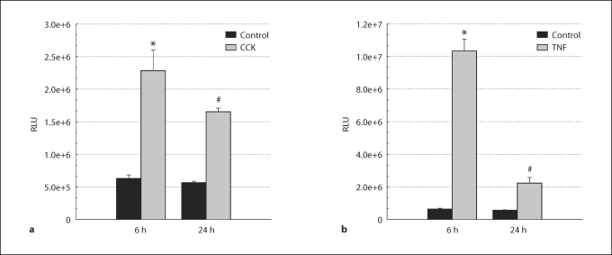
Methods: AR42J cells incorporating an NF-kappaB-responsive luciferase reporter, with and without adenoviral transduction of DNp38, were stimulated with cholecystokinin (CCK) or tumor necrosis factor-alpha (TNF-alpha) prior to measuring NF-kappaB activation.

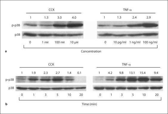
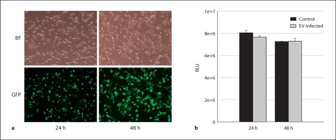
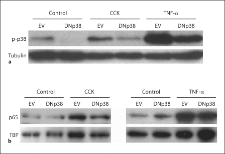
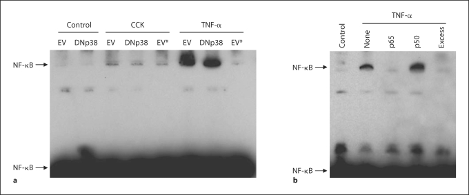
Results: CCK- or TNF-alpha-stimulated NF-kappaB-dependent gene transcription (luciferase assay) was substantially subdued by DNp38 expression. These findings were confirmed by electrophoretic mobility shift assay. Nuclear translocation of the p65 NF-kappaB subunit following agonist stimulation was evident (supershift). Characterization studies showed excellent adenoviral infection efficiency and cell viability in our AR42J cell model. Agonist-stimulated dose- and time-dependent p38 activation, with inhibition by DNp38 expression, was also confirmed.

Conclusion: The p38 MAP kinase regulates NF-kappaB pathway activation in exocrine pancreatic cells, and thus potentially plays a role in the mechanism of acute pancreatitis pathogenesis..


Figures

Fig. 1
Fig. 1
Dose response and time course of p38 activation in AR42J cells following CCK or TNF-α stimulation. a Cells were stimulated with increasing concentrations of either CCK or TNF-α for 5 min and immunoblots performed using antibody specific to phosphorylated p38. Dose-dependent p38 activation following CCK stimulation occurred at 100-nM concentrations and above, while TNF-α stimulated p38 activation occurred at 1-ng/ml doses and above. b AR42J cells were stimulated with 10 μM CCK or 10 ng/ml TNF-α for various periods of time from 0 to 20 min and immunoblots were performed using antibody specific to phosphorylated p38. Time-dependent p38 activation peaked at 5 min following CCK stimulation, and 10 min following TNF-α stimulation. In all panels, densitometry ratios normalized to total p38 expression are provided above each lane with the control value represented as 1.0.
Fig. 2
Fig. 2
Adenovirus infection of AR42J cells. a Cells were infected with 5 MOI of Ad.GFP, then imaged at 24 and 48 h post-infection. Comparison of bright field (BF) filter view (total number of cells) to the FITC filter view (GFP-expressing cells) for the same fields of cells showed close to 90% infection efficiency by 48 h. b AR42J cells were infected with 5 MOI of Ad.EV or left untreated (control), and cell viability evaluated at 24 h and 48 h post-infection by ATP assay. Results show no significant difference in cell viability in Ad.EV-infected cells in comparison to controls at either 24 h or 48 h. ANOVA, p < 0.05; n = 3 wells/group; RLU = relative light units.
Fig. 3
Fig. 3
Effect of DNp38 expression on p38 and NF-κB activation following CCK or TNF-α stimulation in AR42J cells. a Cells were infected with 5 MOI of Ad.EV or Ad.DNp38 for 48 h, then stimulated for 5 min with 10 μM CCK or 10 ng/ml TNF-α. Immunoblots of whole cell lysates were performed using an antibody specific to phosphorylated p38. Densitometry analysis of the immunoblots normalized to tubulin expression shows that p38 is activated following CCK or TNF-α stimulation, and that this activation is attenuated by the expression of DNp38 by 40% in CCK-stimulated cells, and 46% in TNF-α-stimulated cells. b AR42J cells were infected with 5 MOI of Ad.EV or Ad.DNp38 for 48 h, then stimulated for 10 min with either 100 nM CCK or 10 ng/ml TNF-α. Immunoblots of nuclear extracts were performed using an antibody specific to the p65 NF-κB subunit. Densitometry analysis of immunoblots normalized to TBP expression shows that NF-κB is activated following CCK or TNF-α stimulation, and that this activation is attenuated by the expression of DNp38 by 27% in CCK-stimulated cells and 35% in TNF-α-stimulated cells.
Fig. 4
Fig. 4
Effect of DNp38 expression on NF-κB binding following CCK or TNF-α stimulation in AR42J cells. a Cells were infected for 48 h with 5 MOI Ad.EV or Ad.DNp38, then stimulated for 10 min with 10 μM CCK or 10 ng/ml TNF-α. EMSA immunoblot analysis of nuclear extracts shows increased formation of NF-κB DNA:protein complexes following stimulation with either CCK or TNF-α. Expression of DNp38 prior to stimulation attenuated NF-κB DNA:protein complex formation in comparison to Ad.EV-infected cells. Control reactions where 200-fold excess unlabeled NF-κB DNA was added prior to addition of NF-κB-biotin DNA, denoted by an asterisk (∗), show decreased DNA:protein interaction in comparison to the addition of NF-κB-biotin DNA alone. b AR42J cells were stimulated for 10 min with 10 ng/ml TNF-α. Nuclear extracts were incubated with 2 μg primary antibody specific to NF-κB subunits p50 or p65 prior to the addition of the DNA probe to the EMSA-binding reaction. Immunoblot analysis shows that the addition of antibody specific to the p65 NF-κB subunit interacted with activated NF-κB protein as seen by a loss of the shifted NF-κB DNA:protein complex, while antibody specific to the p50 NF-κB subunit did not diminish or supershift the band. Additionally, there was no formation of specific NF-κB DNA:protein complexes in either the unstimulated control lane or the lane containing a 200-fold excess of unlabeled NF-κB DNA probe.
Fig. 5
Fig. 5
Time course of CCK- or TNF-α-induced NF-κB-dependent gene transcription in AR42J cells. Cells infected with 5 MOI Ad.NF-κB-luc were stimulated with 10 μM CCK or 10 ng/ml TNF-α for 6 or 24 h prior to harvesting 48 h post-infection. a CCK-stimulated cells showed a 3.6-fold increase in NF-κB transcriptional activity after 6 h compared to unstimulated controls, which dropped to a 2.9-fold increase at 24 h. b TNF-α-stimulated cells showed a 16.4-fold increase in luciferase activity at 6 h compared to unstimulated controls, which dropped to a 3.9-fold increase in activity at 24 h. ∗ Significant difference from the unstimulated empty vector control group, ANOVA, p <0.05. # Significant difference from the stimulated empty vector group, paired t test, p <0.05. n = 3 wells per experimental group; RLU = relative light units.
Fig. 6
Fig. 6
Effect of DNp38 expression on NF-κB-dependent gene transcription in AR42J cells stimulated with CCK or TNF-α. Cells were co-infected with 5 MOI of Ad.NF-κB-luc and also 5 MOI of either Ad.EV or Ad.DNp38 for 48 h, then stimulated with 10 μM CCK or 10 ng/ml TNF-α for 6 h prior to harvest. a Cells stimulated with CCK showed a 4.3-fold increase in luciferase activity in Ad.EV-infected cells compared to unstimulated controls, which then decreased by 68% in DNp38-expressing cells. b Cells stimulated with TNF-α exhibited a 13.1-fold increase in transcriptional activity in Ad.EV-infected cells in comparison to unstimulated controls, which decreased by 54% in DNp38-expressing cells. ANOVA, p <0.05. ∗ Significant difference from the unstimulated empty vector control group. # Significant difference from the stimulated empty vector group. n = 3 wells per experimental group; RLU = relative light units.

Similar articles

Cited by

References

    1. Norman J. The role of cytokines in the pathogenesis of acute pancreatitis. Am J Surg. 1998;175:76–83. - PubMed
    1. Fleischer F, Dabew R, Goke B, Wagner AC. Stress kinase inhibition modulates acute experimental pancreatitis. World J Gastroenterol. 2001;7:259–265. - PMC - PubMed
    1. Satoh A, Shimosegawa T, Fujita M, Kimura K, Masamune A, Koizumi M, Toyota T. Inhibition of nuclear factor-κB activation improves the survival of rats with taurocholate pancreatitis. Gut. 1999;44:253–258. - PMC - PubMed
    1. Chen X, Ji B, Han B, Ernst SA, Simeone D, Logsdon CD. NF-κB activation in pancreas induces pancreatic and systemic inflammatory response. Gastroenterology. 2002;122:448–457. - PubMed
    1. Han B, Ji B, Logsdon CD. CCK independently activates intracellular trypsinogen and NF-κB in rat pancreatic acinar cells. Am J Physiol. 2001;280:C465–C472. - PubMed

Publication types