

Diffusion tensor imaging: a review for pediatric researchers and clinicians
- PMID: 20453582
- PMCID: PMC4245082
- DOI: 10.1097/DBP.0b013e3181dcaa8b
Diffusion tensor imaging: a review for pediatric researchers and clinicians
Abstract
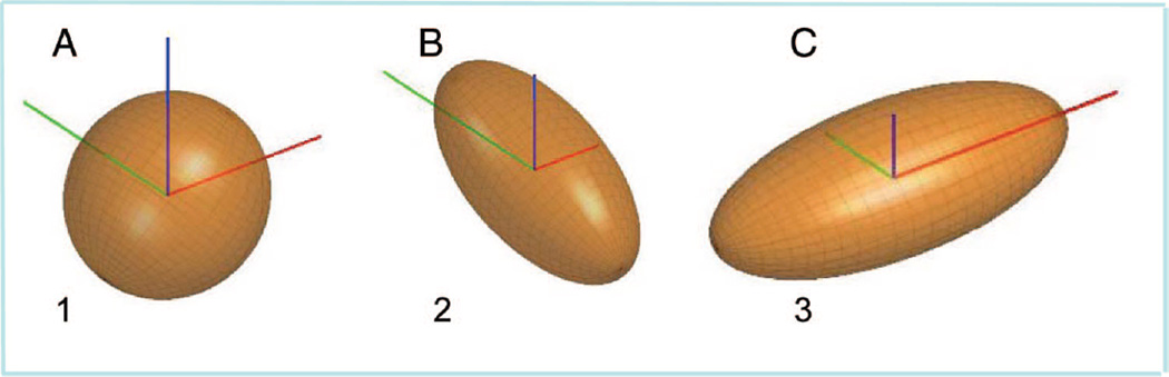
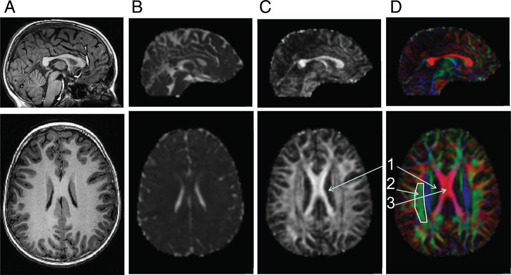
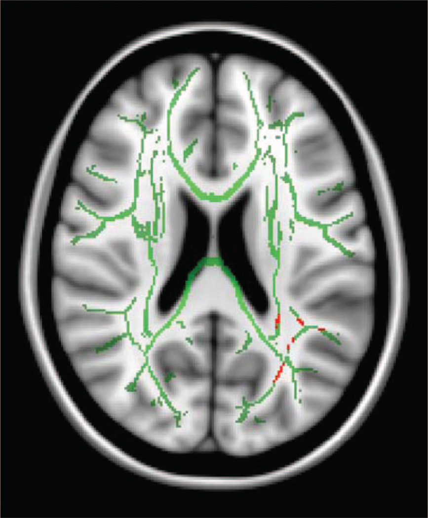
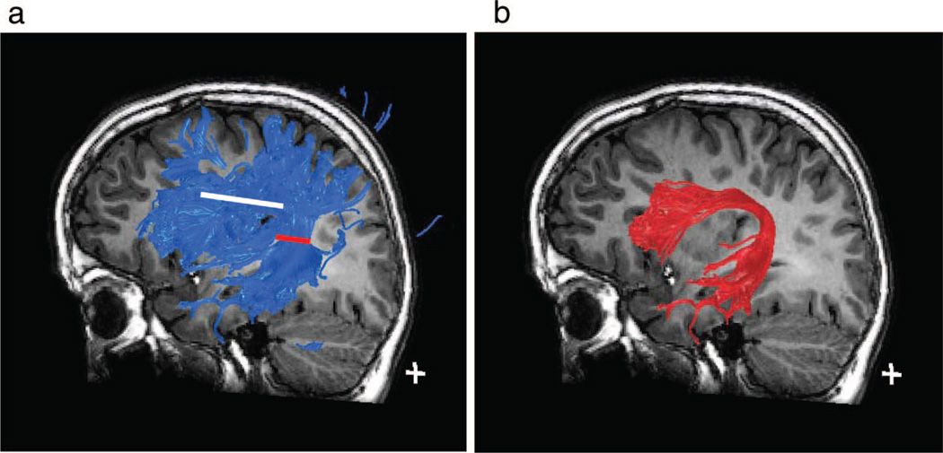
Diffusion tensor imaging (DTI) is a magnetic resonance imaging technique that allows for the visualization and characterization of the white matter tracts of the brain in vivo. DTI does not assess white matter directly. Rather, it capitalizes on the fact that diffusion is isotropic (equal in all directions) in cerebral spinal fluid and cell bodies but anisotropic (greater in one direction than the other directions) in axons that comprise white matter. It provides quantitative information about the degree and direction of water diffusion within individual units of volume within the magnetic resonance image, and by inference, about the integrity of white matter. Measures from DTI can be applied throughout the brain or to regions of interest. Fiber tract reconstruction, or tractography, creates continuous 3-dimensional tracts by sequentially piecing together estimates of fiber orientation from the direction of diffusion within individual volume units. DTI has increased our understanding of white matter structure and function. DTI shows nonlinear growth of white matter tracts from childhood to adulthood. Delayed maturation of the white matter in the frontal lobes may explain the continued growth of cognitive control into adulthood. Relative to good readers, adults and children who are poor readers have evidence of white matter differences in a specific region of the temporo-parietal lobe, implicating differences in connections among brain regions as a factor in reading disorder. Measures from DTI changed in poor readers who improved their reading skills after intense remediation. DTI documents injury to white matter tracts after prematurity. Measures indicative of white matter injury are associated with motor and cognitive impairment in children born prematurely. Further research on DTI is necessary before it can become a routine clinical procedure.
Figures
References
-
- Just MA, Varma S. The organization of thinking: what functional brain imaging reveals about the neuroarchitecture of complex cognition. Cogn Affect Behav Neurosci. 2007;7:153–191. - PubMed
-
- Thomas MSC, McClelland JL. Connectionist Models of Cognition. New York, NY: Cambridge University Press; 2008.
-
- Rogers TT, McClelland JL. A Parallel Distributed Processing Approach to Semantic Cognition: Applications to Conceptual Development. Mahwah, NJ: Lawrence Erlbaum Associates Publishers; 2005.
-
- Fields RD. White matter matters. Sci Am. 2008;298:42–49. - PubMed
Publication types
MeSH terms
Grants and funding
LinkOut - more resources
Full Text Sources
Other Literature Sources
