. 2010 Oct;18(5):462-72.
doi: 10.1097/PAI.0b013e3181dbfcd2.

Novel monoclonal antibodies against the proximal (carboxy-terminal) portions of MUC16

Affiliations

Novel monoclonal antibodies against the proximal (carboxy-terminal) portions of MUC16

Thapi Dharma Rao et al. Appl Immunohistochem Mol Morphol. 2010 Oct.

Abstract

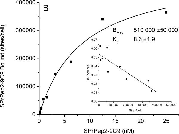
The CA125 antigen, recognized by the OC125 antibody, is a tissue-specific circulating antigen expressed in ovarian cancer. The CA125 antigen is encoded by the MUC16 gene cloned by Yin and Lloyd. The full-length gene describes a complex tethered mucin protein present primarily in a variety of gynecologic tissues, especially neoplasms. OC125 and other related antibodies react with glycosylation-dependent antigens present exclusively in the cleaved portion of the molecule. These antibodies are not useful as screening tools, nor can they detect the proximal residual MUC16 protein fragment after cleavage. This has limited its diagnostic and therapeutic applications. Using synthetic peptides, we raised novel-specific antibodies to the carboxy-terminal portion of MUC16 retained by the cell proximal to the putative cleavage site. These antibodies were characterized using fluorescence-activated cell-sorting analysis, enzyme-linked immunoassay, Western blot analysis, and immunohistochemistry. Each of the selected monoclonal antibodies was reactive against recombinant GST-ΔMUC16 protein and the MUC16-transfected SKOV3 cell line. Three antibodies, 4H11, 9C9, and 4A5 antibodies showed high affinities by Western blot analysis and saturation-binding studies of transfected-SKOV3 cells and displayed antibody internalization. Immunohistochemical positivity with novel antibody 4H11 was similar to OC125 but with important differences, including diffuse positivity in lobular breast cancer and a small percentage of OC125-negative ovarian carcinomas that showed intense and diffuse 4H11. Development of such antibodies may be useful for the characterization of MUC16 biology and allow for future studies in targeted therapy and diagnostics.


Figures

Figure 1
Figure 1
Three MUC16 carboxy terminus peptides were synthesized at the MSKCC Microchemistry Core Facility. Peptide 1 is near the putative cleavage site, peptide 2 is before the transmembrane, and peptide 3 is the internal peptide, which is inside the transmembrane.
Figure 2
Figure 2
Comparison staining of high-grade serous ovarian carcinomas using OC125 (left panel) and 4H11 (right panel)
Figure 3
Figure 3
Immunohistochemical scoring of OC125 and 4H11 on tissue microarrays of high-grade ovarian serous carcinoma. Only membranous and/or cytoplasmic staining was considered positive. Score 0: No staining; Score 1: <5% strong or weak; Score 2: 5–50% strong or weak; Score 3: 51–75% strong or 51–100% weak; Score 4: 76–99% strong; Score 5: 100% strong Figure 3A: OC125 (Score 0); Figure 3B: OC125 (Score 1); Figure 3C: OC125 (Score 2); Figure 3D: OC125 (Score 3); Figure 3E: OC125 (Score 4); Figure 3F: OC125 (Score 5); Figure 3G: 4H11 (Score 0); Figure 3H: 4H11 (Score 1); Figure 3I: 4H11 (Score 2); Figure 3J: 4H11 (Score 3); Figure 3K: 4H11 (Score 4); Figure 3L: 4H11 (Score 5)
Figure 3
Figure 3
Immunohistochemical scoring of OC125 and 4H11 on tissue microarrays of high-grade ovarian serous carcinoma. Only membranous and/or cytoplasmic staining was considered positive. Score 0: No staining; Score 1: <5% strong or weak; Score 2: 5–50% strong or weak; Score 3: 51–75% strong or 51–100% weak; Score 4: 76–99% strong; Score 5: 100% strong Figure 3A: OC125 (Score 0); Figure 3B: OC125 (Score 1); Figure 3C: OC125 (Score 2); Figure 3D: OC125 (Score 3); Figure 3E: OC125 (Score 4); Figure 3F: OC125 (Score 5); Figure 3G: 4H11 (Score 0); Figure 3H: 4H11 (Score 1); Figure 3I: 4H11 (Score 2); Figure 3J: 4H11 (Score 3); Figure 3K: 4H11 (Score 4); Figure 3L: 4H11 (Score 5)
Figure 4A
Figure 4A
Western blot analysis of GST-ΔMUC16c114 fusion protein with monoclonal antibodies 9C9.21.5.13 and 4H11.2.5
Figure 4B
Figure 4B
Western blot analysis of SKOV3-phrGFP-ΔMUC16c114 and SKOV3-phrGFP-ΔMUC16c334 protein extract and probed with monoclonal antibodies 9C9.21.5.13 and 4H11.2.5
Figure 5A
Figure 5A
MUC16 carboxy terminus monoclonal antibodies binding affinity on OVCAR3 cells (Panels A–D)
Figure 5A
Figure 5A
MUC16 carboxy terminus monoclonal antibodies binding affinity on OVCAR3 cells (Panels A–D)
Figure 5A
Figure 5A
MUC16 carboxy terminus monoclonal antibodies binding affinity on OVCAR3 cells (Panels A–D)
Figure 5A
Figure 5A
MUC16 carboxy terminus monoclonal antibodies binding affinity on OVCAR3 cells (Panels A–D)
Figure 5B
Figure 5B
Internalization of radio-labelled 4H11 and OC125 monoclonal antibodies on SKOV3-phrGFP-ΔMUC16c334 stable transfected cells

References

    1. Bast RC, Jr, Feeney M, Lazarus H, et al. Reactivity of a monoclonal antibody with human ovarian carcinoma. J Clin Invest. 1981;68:1331–1337. - PMC - PubMed
    1. Bast RC, Jr, Klug TL, St John E, et al. A radioimmunoassay using a monoclonal antibody to monitor the course of epithelial ovarian cancer. N Engl J Med. 1983;309:883–887. - PubMed
    1. Rustin GJ, Bast RC, Jr, Kelloff GJ, et al. Use of CA-125 in clinical trial evaluation of new therapeutic drugs for ovarian cancer. Clin Cancer Res. 2004;10:3919–3926. - PubMed
    1. Rosen DG, Wang L, Atkinson JN, et al. Potential markers that complement expression of CA125 in epithelial ovarian cancer. Gynecol Oncol. 2005;99:267–277. - PubMed
    1. Bast RC, Jr, Badgwell D, Lu Z, et al. New tumor markers: CA125 and beyond. Int J Gynecol Cancer. 2005;15(Suppl 3):274–281. - PubMed

Publication types

MeSH terms