. 2010 Jun 2;29(11):1928-40.
doi: 10.1038/emboj.2010.84. Epub 2010 May 7.

Deciphering the assembly of the Yersinia type III secretion injectisome

Affiliations

Deciphering the assembly of the Yersinia type III secretion injectisome

Andreas Diepold et al. EMBO J. .

Abstract

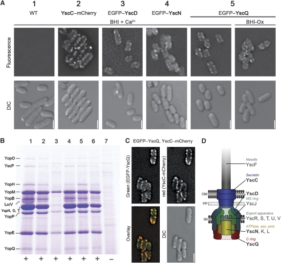
The assembly of the Yersinia enterocolitica type III secretion injectisome was investigated by grafting fluorescent proteins onto several components, YscC (outer-membrane (OM) ring), YscD (forms the inner-membrane (IM) ring together with YscJ), YscN (ATPase), and YscQ (putative C ring). The recombinant injectisomes were functional and appeared as fluorescent spots at the cell periphery. Epistasis experiments with the hybrid alleles in an array of injectisome mutants revealed a novel outside-in assembly order: whereas YscC formed spots in the absence of any other structural protein, formation of YscD foci required YscC, but not YscJ. We therefore propose that the assembly starts with YscC and proceeds through the connector YscD to YscJ, which was further corroborated by co-immunoprecipitation experiments. Completion of the membrane rings allowed the subsequent assembly of cytosolic components. YscN and YscQ attached synchronously, requiring each other, the interacting proteins YscK and YscL, but no further injectisome component for their assembly. These results show that assembly is initiated by the formation of the OM ring and progresses inwards to the IM ring and, finally, to a large cytosolic complex.


Conflict of interest statement

The authors declare that they have no conflict of interest.

Figures

Figure 1
Figure 1
Fluorescently labelled Ysc proteins are functional and allow visualization of the injectisome. (A) Fluorescence deconvolution microscopy showing the formation of fluorescent spots at the bacterial membrane of Y. enterocolitica bacteria grown in secretion-non-permissive (BHI+Ca2+) and secretion-permissive medium (BHI-Ox): 1—E40(pYV40) [wild type], 2—E40(pMA4005) [YscC–mCherry], 3—E40(pAD4050) [EGFP–YscD], 4—E40(pAD4136)(pAD182) [ΔYscN+pBAD–egfp–yscN], 5—E40(pAD4016) [EGFP–YscQ]. All fusion proteins except for EGFP–YscN are encoded under their native promoter on the pYV virulence plasmid. Upper lane: mCherry fluorescence for strain 2, EGFP fluorescence for other strains; lower lane: corresponding DIC picture. All fluorescence pictures were taken 3 h after the induction of the T3S system by temperature shift to 37°C. Scale bars: 2 μm. (B) Analysis of the Yop proteins secreted in secretion-permissive conditions. The tagged strains are fully functional for effector secretion, except for the strain expressing EGFP–YscD (lane 3), which shows reduced secretion. Culture supernatants were separated on a 12% SDS–PAGE gel and stained with Coomassie Brilliant Blue. Strains as listed in (A), 6—E40(pMAAD4006) [EGFP–YscQ, YscC–mCherry], 7—E40(pAD4051) [ΔYscD, negative control]. Bottom line: Needle formation (+/−) in the tested strains (data not shown). (C) Fluorescence microscopy showing the colocalization of EGFP–YscQ with YscC–mCherry in E40(pMAAD4006) bacteria. Fluorescent pictures were obtained as described in (A). (D) Model of the Yersinia Ysc injectisome. Fluorescently labelled proteins are shown in bold print. OM, outer membrane; PP, periplasm; IM, inner membrane.
Figure 2
Figure 2
YscC assembly only requires its pilotin; YscD assembly requires the presence of YscC, but not of YscJ. Copurification of the three structural ring proteins suggests the stepwise assembly order YscC–YscD–YscJ. (A) Fluorescence microscopy showing the formation of secretin spots [YscC–mCherry] at the bacterial membrane in a strain lacking the virulence plasmid pYV, after in trans expression of YscC–mCherry and YscW (plasmids pMA8, pRS6) for 3 h at 37°C. Scale bars: 2 μm. (B) Fluorescence microscopy showing the formation of YscD spots at the bacterial membrane in strains E40(pAD4050) [EGFP–YscD], E40(pMAAD4018) [EGFP–YscD, ΔYscC], and E40(pAD4080) [EGFP–YscD, ΔYscJ]. YscD remains cytosolic in the absence of YscC, whereas it assembles in membrane spots in the absence of YscJ. (C) Analysis of the copurification of YscC and YscD after affinity purification of YscJ. Deletion of either YscC or YscD abolishes the copurification of the respective other protein with YscJ-FLAG-His. Bacteria were incubated for 3 h at 37°C, spheroplasted, and lysed. Proteins were purified by FLAG affinity, separated on 4–12% gradient SDS–PAGE, and analysed by immunoblot with the respective anti-YscC, -YscD, or -YscJ antibodies. All strains were ΔYadA to facilitate cell lysis: 1—E40(pLJM4029) [WT], 2—E40(pAD4054) [YscJ-FLAG-His], 3—E40(pAD4109) [YscJ-FLAG-His, ΔYscC], 4—E40(pAD4110) [YscJ-FLAG-His, ΔYscD], 5—E40(pAD4112) [YscJ-FLAG-His, ΔYscQ]. (D) Analysis of the copurification of YscC and YscJ after affinity purification of YscD. Whereas deletion of YscC abolishes copurification of YscJ with His-FLAG-YscD, YscJ is not required for the interaction between YscC and YscD. Samples were obtained as described for (C). All strains were ΔYadA to facilitate cell lysis: 1—E40(pLJM4029) [WT], 2—E40(pAD4055) [His-FLAG-YscD], 3—E40(pADMA4101) [His-FLAG-YscD, ΔYscC], 4—E40(pAD4089) [His-FLAG-YscD, ΔYscJ].
Figure 3
Figure 3
C ring formation requires both transmembrane rings and the ATPase complex, but not the export apparatus or any secreted substrate. (A) Fluorescence microscopy pictures of bacteria expressing EGFP–YscQ combined with deletions of different genes. Micrographs were taken 3 h after induction of the T3S system. (For the control strains and the strains with deletions in the proteins required for C ring formation—upper lane: EGFP fluorescence, lower lane: corresponding DIC picture). Scale bars: 2 μm. (B) Schematic representation of the injectisome showing the components required (bold, dark) and not required (normal, light) for C ring formation. For information about the used strains, refer to Table I.
Figure 4
Figure 4
The assembly of the ATPase requires both transmembrane rings, YscK, YscL, and YscQ, but not the export apparatus. Fluorescence microscopy pictures of YscN null mutants complemented with EGFP–YscN combined with deletions of different genes. Wild-type protein levels were established by EGFP–YscN induction with 0.05% arabinose. Micrographs were taken 3 h after induction of EGFP–YscN and the T3S system. Scale bars: 2 μm. For information about the used strains, refer to Table I.
Figure 5
Figure 5
The structure, but not the ATPase activity of YscN, is required for the formation of the C ring. (A) Fluorescence microscopy showing the formation of C ring spots in E40(pAD4104) [EGFP–YscQ, ΔYscN], complemented with plasmids encoding wild-type YscN or the catalytically inactive YscN(K175E), 3 h after induction of the type III secretion system. Scale bars: 2 μm. (B) Upper part: Analysis of Yop protein secretion in strain E40(pAD4104) complemented with wild type, or catalytically inactive YscN. Expression of YscN was either not induced (−) or induced with 0.05% arabinose (+). Culture supernatants were separated on a 12% SDS–PAGE gel and stained with Coomassie Brilliant Blue. Lower part: Expression of YscN in the corresponding strains. Cell pellets were separated on a 12% SDS–PAGE gel and analysed by immunoblot with anti-YscN antibodies. (C) Export of different classes of substrates in strains expressing EGFP–YscQ, and the different YscN variants. Culture supernatants were separated on a 15% SDS–PAGE gel and analysed by immunoblot with the respective antibodies.
Figure 6
Figure 6
Formation of the C ring occurs about 60 min after induction of the T3S system. It directly precedes needle formation and effector secretion. (A) Fluorescence microscopy showing the formation of C ring spots [EGFP–YscQ] in strain E40(pAD4016) at various time points after induction of the synthesis of the T3S system by temperature shift to 37°C (upper lane: EGFP fluorescence; lower lane: corresponding DIC picture). Scale bars: 2 μm. (B) Time course of needle formation in wild-type strain E40(pYV40). Needle formation was monitored by SDS–PAGE analysis of purified needles. The needle pellet was separated on a 15% gel and analysed by immunoblot with anti-LcrV and anti-YscF antibodies, respectively. (C) Time course of Yop protein secretion by wild-type strain E40(pYV40). Culture supernatants were separated on a 12% SDS–PAGE gel and stained with Coomassie Brilliant Blue. All time-course experiments were performed in secretion-permissive conditions (Ca2+-depleted medium).
Figure 7
Figure 7
Model of assembly of the Yersinia injectisome. Formation of the injectisome is initiated by formation of the secretin ring in the outer membrane. Next, YscD attaches to YscC, which allows the subsequent completion of the MS ring by attachment of YscJ. After the formation of the membrane ring structures, the ATPase–C ring complex, consisting of YscN, K, L, and Q assembles at the cytoplasmic side of the injectisome. The exact time point of the integration of the IM proteins YscR, S, T, U, and V is unclear. Afterwards, the needle consisting of YscF and LcrV can be assembled. Bold font denotes protein names, normal font denotes functional subunits. The global structure of YscC, D, J is derived from Spreter et al (2009.

Similar articles

Cited by

References

    1. Abrahams JP, Leslie AG, Lutter R, Walker JE (1994) Structure at 2.8 A resolution of F1-ATPase from bovine heart mitochondria. Nature 370: 621–628 - PubMed
    1. Akeda Y, Galan JE (2005) Chaperone release and unfolding of substrates in type III secretion. Nature 437: 911–915 - PubMed
    1. Alfano JR, Collmer A (2004) Type III secretion system effector proteins: double agents in bacterial disease and plant defense. Annu Rev Phytopathol 42: 385–414 - PubMed
    1. Blaylock B, Riordan KE, Missiakas DM, Schneewind O (2006) Characterization of the Yersinia enterocolitica type III secretion ATPase YscN and its regulator, YscL. J Bacteriol 188: 3525–3534 - PMC - PubMed
    1. Blocker A, Gounon P, Larquet E, Niebuhr K, Cabiaux V, Parsot C, Sansonetti P (1999) The tripartite type III secreton of Shigella flexneri inserts IpaB and IpaC into host membranes. J Cell Biol 147: 683–693 - PMC - PubMed

Publication types

MeSH terms

Substances