. 2010 Jun;17(6):753-60.
doi: 10.1038/nsmb.1816. Epub 2010 May 9.

p53 activates transcription by directing structural shifts in Mediator

Affiliations

p53 activates transcription by directing structural shifts in Mediator

Krista D Meyer et al. Nat Struct Mol Biol. 2010 Jun.

Abstract

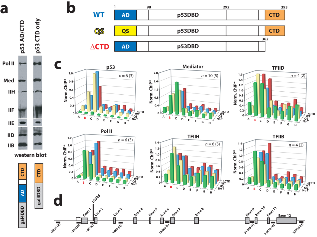
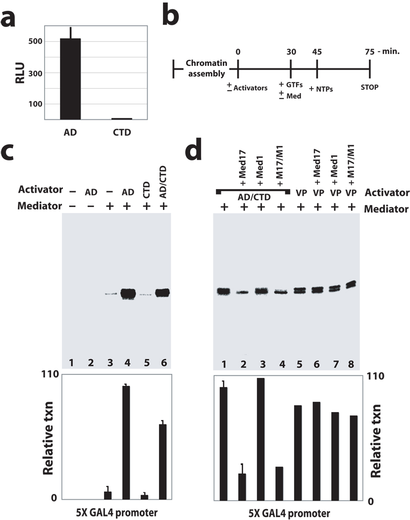
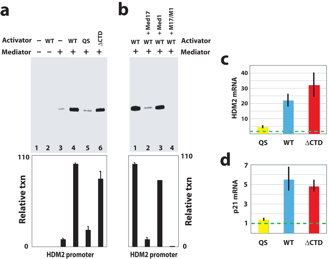
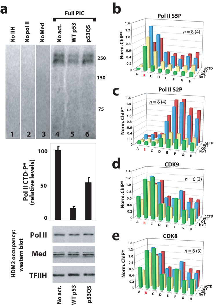
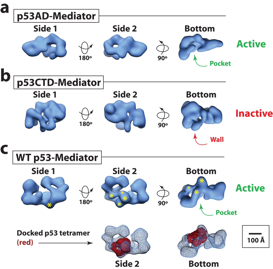
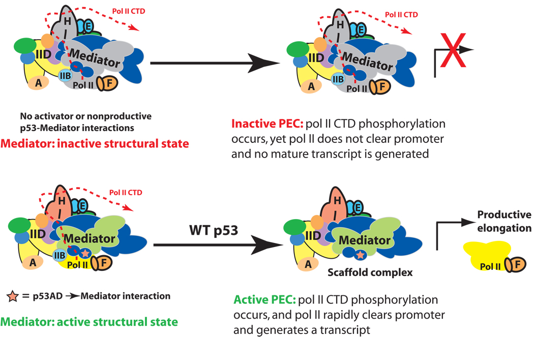
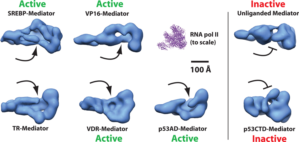
It is not well understood how the human Mediator complex, transcription factor IIH and RNA polymerase II (Pol II) work together with activators to initiate transcription. Activator binding alters Mediator structure, yet the functional consequences of such structural shifts remain unknown. The p53 C terminus and its activation domain interact with different Mediator subunits, and we find that each interaction differentially affects Mediator structure; strikingly, distinct p53-Mediator structures differentially affect Pol II activity. Only the p53 activation domain induces the formation of a large pocket domain at the Mediator-Pol II interaction site, and this correlates with activation of stalled Pol II to a productively elongating state. Moreover, we define a Mediator requirement for TFIIH-dependent Pol II C-terminal domain phosphorylation and identify substantial differences in Pol II C-terminal domain processing that correspond to distinct p53-Mediator structural states. Our results define a fundamental mechanism by which p53 activates transcription and suggest that Mediator structural shifts trigger activation of stalled Pol II complexes.


Figures

Figure 1
Figure 1
The p53AD is not required for PEC assembly in vitro or in cells. (a) Immobilized template assays. Occupancy of PEC components in the presence of promoter-bound GAL4–p53CTD or a GAL4–p53AD/CTD fusion protein. Note that endogenous p53 will not bind the GAL4 promoter. Antibodies—pol II: Rpb1; Mediator: Med1; IIH: ERCC3; IIF: Rap74; IIE: IIEα; IID: TBP. (b) Schematic of WT p53 and mutant p53 proteins used in this study. (c) ChIP assays at HDM2 in p53-null HCT116 cells following transfection with wild-type p53 (WT, blue bars), p53 with a truncated CTD (residues 1–362; ΔCTD, red bars), or p53QS mutant (QS, yellow bars). Data from no transfection controls (No T, green bars) are also shown. The probe location in red (B) represents the promoter/transcription start site. Note that PEC factors appear to be pre-loaded at the promoter, as observed previously. *ChIP output was normalized to WT p53, typically from C primer. For clarity, error bars are not shown but can be viewed in Supplementary Figure 5. (d) Schematic of the HDM2 locus, showing ChIP probe locations.
Figure 2
Figure 2
p53AD is required to activate stalled pol II in vitro and in cells; PECs assembled in the absence of p53 or with p53CTD remain inactive. (a) The p53CTD cannot activate transcription in cells. Luciferase reporter assays in HEK293 cells transfected with GAL4–p53AD or GAL4–p53CTD; results shown represent mean and standard error from three independent experiments. (b) Outline of reconstituted transcription assay. (c) Reconstituted transcription on a promoter containing tandem GAL4 sites; AD: GAL4–p53AD; CTD: GAL4–p53CTD; AD/CTD: GAL4–p53AD/CTD fusion protein. Representative data are shown; plot shows mean and s.e.m. from multiple experiments (n = 4, 10, 6, 4 for lanes 3–6). (d) Recombinant Med17, which blocks the p53AD–Mediator interaction, can block p53-dependent activation in vitro. Transcription is repressed upon addition of recombinant Med17 (lanes 2 and 4); however, Med17 had no detectable impact on VP16-dependent activation (lanes 5–8). VP16 interacts with Mediator through the Med25 subunit,. Quantitation of transcripts is shown in the bar graph, and bars represent the s.e.m. for lanes 1 and 2 (n = 10, 2, respectively). Each experiment shown in c and d contained equivalent amounts of chromatin template, general transcription factors (TFIIA, IIB, IID, IIE, IIF, IIH, and pol II), and NTPs. Activators and Mediator added as shown.
Figure 3
Figure 3
p53AD activates stalled pol II within the context of the native tetramer in vitro and in cells. (a) Reconstituted transcription from the native HDM2 promoter. As with the GAL4 template, transcription was dependent upon Mediator and an intact p53AD. Activators tested were wild-type p53 (WT), p53 with a mutated activation domain (QS), and p53 lacking the CTD (ΔCTD). Each p53 protein was purified as a tetramer. Plot shows mean and s.e.m. from multiple experiments (n = 5, 10, 4, 4 for lanes 3–6). (b) As with the GAL4 template (Fig. 2d), recombinant Med17 could strongly repress p53-dependent transcription at the HDM2 promoter, whereas recombinant Med1 had little effect. The bar graph shows quantitation of transcripts, with bars representing s.e.m. (n = 10, 2 for lanes 1 and 2). Experiments shown in a and b followed the same timeline as shown in Figure 2b and contained the same concentration of HDM2 chromatin template, TFIIA, IIB, IID, IIE, IIF, IIH, pol II, and NTPs. Mediator and activators added as shown. (c) and (d) Summary of results from RT-QPCR experiments measuring HDM2 or p21 mRNA levels in response to the p53 proteins shown. Experiments were completed in p53-null HCT116 cells; dashed line represents mRNA levels measured in non-transfected control cells. Note that similar mRNA levels observed with WT p53 and p53ΔCTD result from their overexpression upon transfection. When expressed at physiological levels, p53ΔCTD is still observed to activate HDM2 and p21, but to a lesser extent relative to WT p53, due to its decreased ability to stably bind DNA.
Figure 4
Figure 4
Mediator is required for TFIIH-dependent pol II CTD phosphorylation within the PEC; oncogenic mutations within p53AD (p53QS) prevent activation of stalled pol II. (a) Immobilized template assays with the native HDM2 promoter. Purified PEC factors (TFIIA, IIB, IID, IIE, IIF, IIH, Mediator, and pol II; see ref. 20) were allowed to assemble on the HDM2 promoter; unbound factors were removed by washing, at which time rNTPs were added (including [32P]-ATP) to visualize and quantitate pol II CTD phosphorylation. Data shown follows a 6-minute incubation with rNTPs; radio-labeled bands migrated around 250 kDa, the approximate size of Rpb1. Bar graph represents mean and s.e.m. from multiple independent experiments (n = 6, 10, 4 for lanes 4–6, respectively). Occupancy of PEC components was measured by western blot, several of which are shown below the bar graph. PECs lacking only one specific component (TFIIH, pol II, or Mediator: lanes 1–3) indicate a Mediator requirement for pol II CTD phosphorylation. (b) and (c) ChIP data reveals that phosphorylated forms of pol II are diminished within the HDM2 gene in the absence of an intact p53AD. (d) and (e) Occupancy of pol II CTD kinases CDK9 (a component of P-TEFb) and CDK8 are not negatively impacted by p53AD mutations. Note that occupancy of another major pol II CTD kinase, TFIIH, is also not altered by p53AD mutations, as observed by ChIP in Figure 1c. The probe location in red (B) represents the promoter/transcription start site. *ChIP output was normalized to WT p53, typically from primer C. For clarity, error bars are not shown but can be viewed in Supplementary Figure 8.
Figure 5
Figure 5
p53AD triggers a structural shift within Mediator distinct from p53CTD. (a) and (b) Different views of Mediator bound to p53AD or p53CTD. Volumes shown at 34 Å and 38 Å resolution, respectively, are rendered to 1.2 MDa, the approximate molecular weight of each complex. A defined pocket domain is evident in p53AD–Mediator (transcriptionally active) whereas in p53CTD–Mediator (transcriptionally inactive), this region is occupied by a wall of protein density. Bar: 100 Å. Note the pocket domain corresponds to the Mediator-pol II interaction site, as shown in Supplementary Figure 14c. (c) The structure of Mediator bound to the wild-type p53 tetramer (34 Å resolution). Like p53AD–Mediator, the pocket domain represents a prominent structural feature. The volume shown is rendered to 1.4 MDa, the approximate molecular weight of the WT p53–Mediator assembly. The general location of the p53 tetramer is highlighted, with asterisks representing the p53 DNA-binding domains. Larger asterisks are closer to the viewer. Lower panels show views of the docked WT p53 tetramer structure within the WT p53–Mediator EM map. The p53 tetramer is solid whereas WT p53–Mediator is shown in mesh.
Figure 6
Figure 6
Model outlining how p53AD appears to trigger pol II promoter escape and transcription elongation. Top: Despite full PEC assembly, promoter-proximally stalled pol II remains unable to transition to a productively elongating state in the absence of the p53AD-directed structural shift within Mediator. Bottom: Mediator structural shifts, orchestrated by p53AD, activate stalled pol II to transition to a productively-elongating state. This Mediator-dependent activation appears to be triggered by formation of the pocket domain within Mediator, which interfaces extensively with pol II. Note the location and path of the pol II CTD within the PEC has not been clearly defined.
Figure 7
Figure 7
The pocket domain represents a common structural feature within activator-bound Mediator. Shown are “bottom” views of Mediator complexes bound to different activation domains. The location of the pocket is noted by the arrows. Mediator structures labeled “active” have been shown to activate transcription in a reconstituted system. Note that transcriptionally inactive structures (unliganded Mediator or p53CTD–Mediator structure) lack the pocket domain. The unliganded Mediator structure is not bound to an activator. Studies with both yeast and human Mediator (Supplementary Fig. 14c) reveal that the pocket domain represents the pol II binding site within Mediator. For reference, the structure of the yeast pol II enzyme (pdb 1y1v) is also shown to the same relative scale.

Similar articles

Cited by

References

    1. Holstege FC, et al. Dissecting the regulatory circuitry of a eukaryotic genome. Cell. 1998;95:717–728. - PubMed
    1. Fondell JD, Ge H, Roeder RG. Ligand induction of a transcriptionally active thyroid hormone receptor coactivator complex. Proc. Natl. Acad. Sci. USA. 1996;93:8329–8333. - PMC - PubMed
    1. Esnault C, et al. Mediator-dependent recruitment of TFIIH modules in Preinitiation Complex. Mol Cell. 2008;31:337–346. - PubMed
    1. Davis JA, Takagi Y, Kornberg RD, Asturias FA. Structure of the yeast RNA polymerase II holoenzyme: Mediator conformation and polymerase interaction. Mol Cell. 2002;10:409–415. - PubMed
    1. Johnson KM, Wang J, Smallwood A, Arayata C, Carey M. TFIID and human mediator coactivator complexes assemble cooperatively on promoter DNA. Genes & Development. 2002;16:1852–1863. - PMC - PubMed

Publication types

MeSH terms