

Tribulosin protects rat hearts from ischemia/reperfusion injury
- PMID: 20453871
- PMCID: PMC4002968
- DOI: 10.1038/aps.2010.45
Tribulosin protects rat hearts from ischemia/reperfusion injury
Abstract
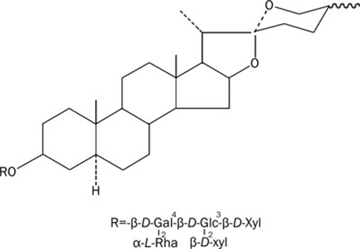
Aim: To investigate the protective effect of tribulosin, a monomer of the gross saponins from Tribulus terrestris, against cardiac ischemia/reperfusion injury and the underlying mechanism in rats.
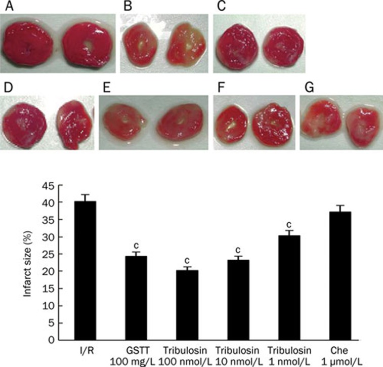
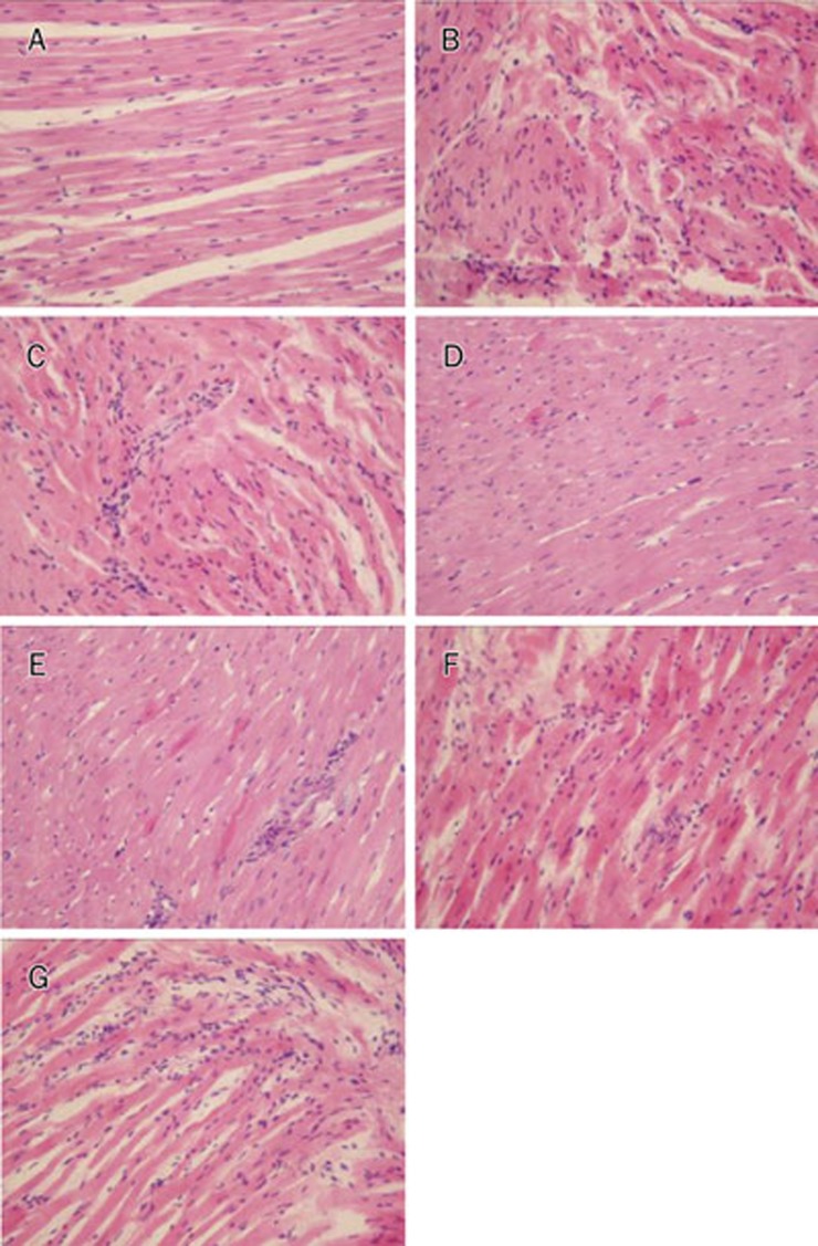
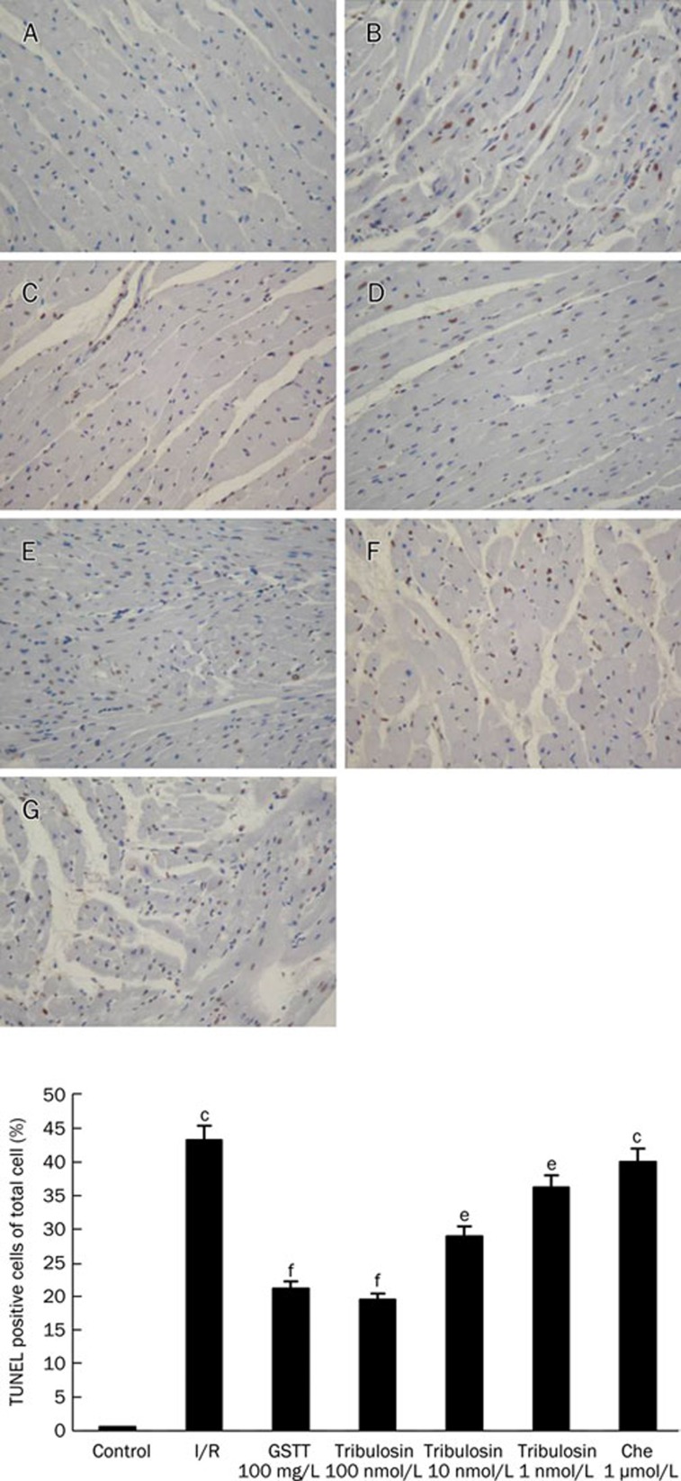
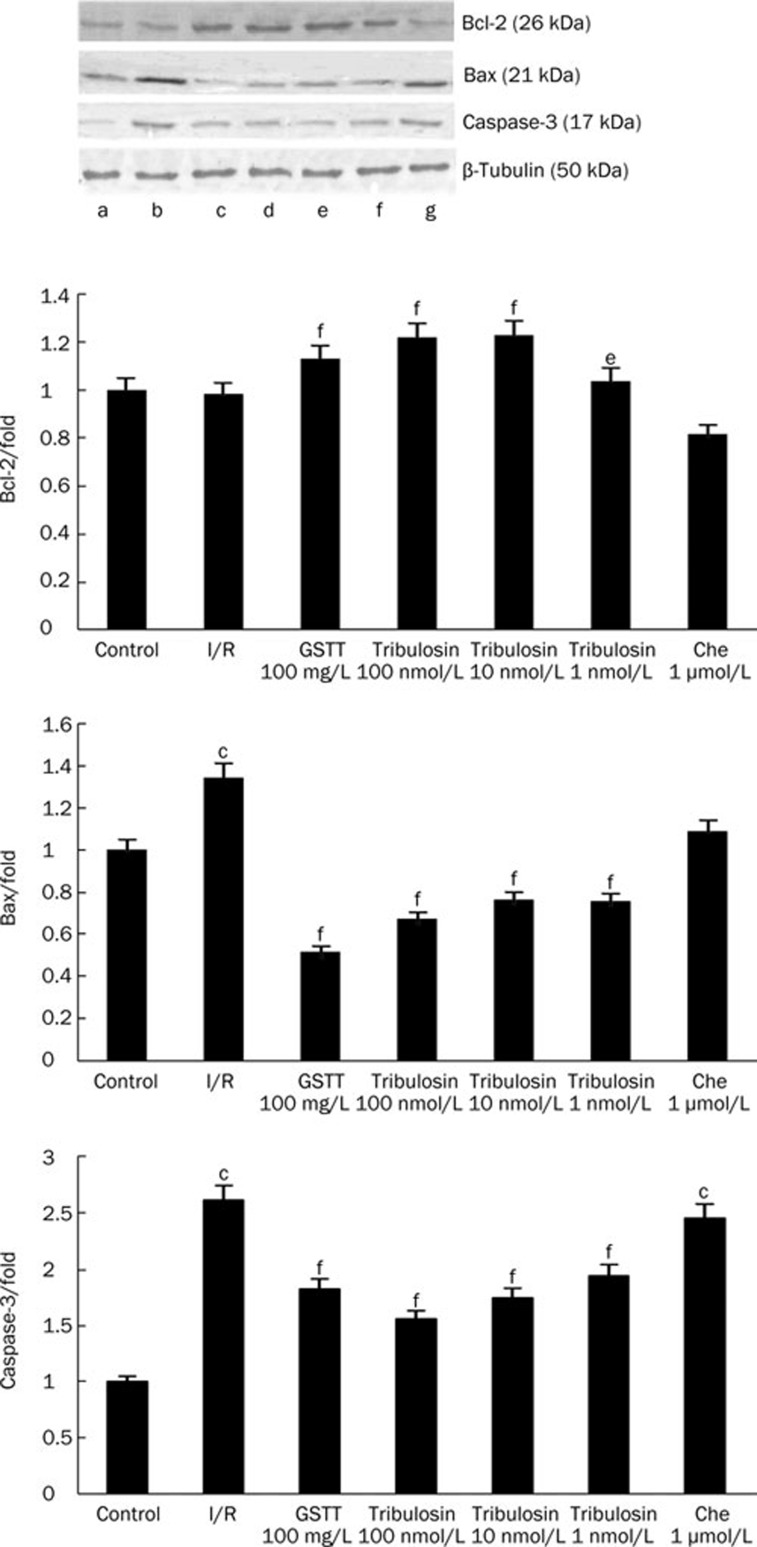
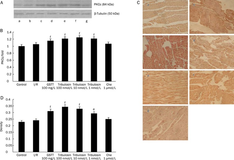
Methods: Isolated rat hearts were subjected to 30 min of ischemia followed by 120 min of reperfusion using Langendorff's technique. The hearts were assigned to seven groups: control, ischemia/reperfusion (I/R), treatment with gross saponins from Tribulus terrestris (GSTT) 100 mg/L, treatment with tribulosin (100, 10, and 1 nmol/L) and treatment with a PKC inhibitor (chelerythrine) (1 micromol/L). Infarct size was assessed by triphenyltetrazolium chloride staining. Malondialdehyde (MDA), aspartate aminotransferase (AST), and lactate dehydrogenase (LDH) contents as well as superoxide dismutase (SOD) and creatine kinase (CK) activities were determined after the treatment. Histopathological changes in the myocardium were observed using hematoxylin-eosin (H&E) staining. Apoptosis was detected with terminal deoxynucleotidyl transferase nick-end labeling (TUNEL) assay. Bcl-2, Bax, caspase-3, and PKCepsilon protein expression were examined using Western blotting.
Results: Tribulosin treatment significantly reduced MDA, AST, CK and LDH contents, and increased the activity of SOD. The infarct size of I/R group was 40.21% of the total area. GSTT and various concentrations of tribulosin treatment decreased the infarct size to 24.33%, 20.24%, 23.19%, and 30.32% (P<0.01). Tribulosin treatment reduced the myocardial apoptosis rate in a concentration-dependent manner. Bcl-2 and PKCepsilon protein expression was increased after tribulosin preconditioning, whereas Bax and caspase-3 expression was decreased. In the chelerythrine group, Bcl-2 and PKCepsilon expression was decreased, whereas Bax and caspase-3 expression was increased.
Conclusion: Tribulosin protects myocardium against ischemia/reperfusion injury through PKCepsilon activation.
Figures
Similar articles
-
[Mechanisms of gross saponins of Tribulus terrestris via activating PKCepsilon against myocardial apoptosis induced by oxidative stress].Yao Xue Xue Bao. 2009 Feb;44(2):134-9. Yao Xue Xue Bao. 2009. PMID: 19408681 Chinese.
-
Influences of remifentanil on myocardial ischemia-reperfusion injury and the expressions of Bax and Bcl-2 in rats.Eur Rev Med Pharmacol Sci. 2018 Dec;22(24):8951-8960. doi: 10.26355/eurrev_201812_16665. Eur Rev Med Pharmacol Sci. 2018. PMID: 30575939
-
Tribulosin suppresses apoptosis via PKC epsilon and ERK1/2 signaling pathway during hypoxia/reoxygenation in neonatal rat ventricular cardiac myocytes.J Asian Nat Prod Res. 2011 Dec;13(12):1135-45. doi: 10.1080/10286020.2011.627327. J Asian Nat Prod Res. 2011. PMID: 22115037
-
[Effects of propofol on cardiomyocytes apoptosis and its mechanism after ischemia/reperfusion injury in isolated rat hearts].Zhongguo Ying Yong Sheng Li Xue Za Zhi. 2008 Feb;24(1):56-61. Zhongguo Ying Yong Sheng Li Xue Za Zhi. 2008. PMID: 21141559 Chinese.
-
Cardioprotective effects of saponins from Panax japonicus on acute myocardial ischemia against oxidative stress-triggered damage and cardiac cell death in rats.J Ethnopharmacol. 2012 Mar 6;140(1):73-82. doi: 10.1016/j.jep.2011.12.024. Epub 2011 Dec 31. J Ethnopharmacol. 2012. PMID: 22226974
Cited by
-
Saponins of Tribulus terrestris attenuated neuropathic pain induced with vincristine through central and peripheral mechanism.Inflammopharmacology. 2019 Aug;27(4):761-772. doi: 10.1007/s10787-018-0502-0. Epub 2018 Jun 25. Inflammopharmacology. 2019. PMID: 29938333
-
Cardiac Protection of Valsartan on Juvenile Rats with Heart Failure by Inhibiting Activity of CaMKII via Attenuating Phosphorylation.Biomed Res Int. 2017;2017:4150158. doi: 10.1155/2017/4150158. Epub 2017 Apr 27. Biomed Res Int. 2017. PMID: 28536695 Free PMC article.
-
Efficacy and safety of 11 oral preparations of single-source traditional Chinese medicines in the treatment of unstable angina pectoris: a systematic review and network meta-analysis.Front Pharmacol. 2025 Jun 24;16:1582661. doi: 10.3389/fphar.2025.1582661. eCollection 2025. Front Pharmacol. 2025. PMID: 40630122 Free PMC article.
-
Anti-Apoptotic Effects of Resistance Training and Tribulus Terrestris Consumption in the Heart Tissue of Rats Exposed to Stanozolol.Eurasian J Med. 2021 Jun;53(2):79-84. doi: 10.5152/eurasianjmed.2021.20051. Eurasian J Med. 2021. PMID: 34177287 Free PMC article.
-
The effects of N-acetylcysteine on cisplatin-induced cardiotoxicity on isolated rat hearts after short-term global ischemia.Toxicol Rep. 2015 Jul 17;2:996-1006. doi: 10.1016/j.toxrep.2015.07.009. eCollection 2015. Toxicol Rep. 2015. PMID: 28962440 Free PMC article.
References
-
- Shi CJ, Qu WJ, Wang JS, Deng TT. Effect of Tribu Saponin from Tribulus terrestris on the formation of atherosclerosis in rats. Nat Prod Res Dev. 2009;21:53–7.
-
- Liu XM, Huang QF, Zhang YL, Lou JL, Liu HS, Zheng H. Effects of Tribulus terrestris L saponion on apoptosis of cortical neurons induced by hypoxia-reoxygenation in rats. J Chin Int Med. 2008;6:45–50. - PubMed
-
- Lu WW, Qu JB, Yang SJ. Influences of GSTT on hemodynamics and oxygen metabolism in anesthetic thoraco-opened dogs. J Jilin Univ (Med Ed) 2006;32:379–82.
-
- Sun W, Li H, Yang SJ. A triterpene saponin from Tribulus terrestris attenuates apoptosis in cardiocyte via activating PKC signalling transduction pathway. J Asian Nat Prod Res. 2008;10:39–48. - PubMed
-
- Mu YL, Xie YY, Zhou L, Zhong Y, Liu L, Bai H, et al. Cardioprotective effect of methylamine irisolidone, a new compound, in hypoxia/reoxygenation injury in cultured rat cardiac myocyte. Chem Biodivers. 2009;6:1170–7. - PubMed
Publication types
MeSH terms
Substances
LinkOut - more resources
Full Text Sources
Other Literature Sources
Medical
Research Materials
